





Expo
ADLM
view channel
view channel
view channel
view channel
view channel
view channel
view channel
view channel
view channel
Clinical Chem.Molecular DiagnosticsHematologyImmunologyMicrobiology
TechnologyIndustry
Events
Webinars

- New Immunoassay Enables Ultrasensitive Blood-Based Tau Tangle Measurement
- Blood Hormone Pattern Distinguishes Endometriosis with High Accuracy
- Blood Test Brings Alzheimer’s Biomarker Assessment to Routine Labs
- Alzheimer’s Biomarkers Identify Faster Cognitive Decline in Adults Over 80
- ADLM Issues Laboratory Guidance for Gender-Diverse Patient Care
- Community-Based Genetic Screening Reaches Rural and Vulnerable Populations
- Blood and Urine Liquid Biopsy Detects Early Colorectal Cancer Mutations
- Epigenetic Profiling Could Refine Prognosis in Acute Myeloid Leukemia
- Genetic Risk Score Supports Diagnosis and Prognosis in Idiopathic Pulmonary Fibrosis
- Extracellular Vesicle Marker Identifies Early Lung Adenocarcinoma and Predicts Recurrence
- AI Decision Support System Guides Treatment Selection for Complex Blood Cancers
- Blood Test Helps Predict Short-Term Mortality After Severe Heart Attack
- Next-Generation Hematology Platform Streamlines High-Complexity Lab Workflows
- Blood Eosinophil Count May Predict Cancer Immunotherapy Response and Toxicity
- Higher Ferritin Threshold May Improve Iron Deficiency Detection in Children
- Anti-Lipid Antibody Biomarkers May Identify Early Lyme Disease and Persistent Symptoms
- Emergency Department Opt-Out Testing Program Identifies Undiagnosed HIV
- Immune Biomarkers Could Identify Risk of Chronic Critical Illness on ICU Admission
- New Cellular Biomarkers Correlate with Disease Severity in Sjögren Disease
- Airway Immune Signature May Predict Tuberculosis Progression Risk
- Gut Microbiome Analysis Identifies Frailty-Related Signatures in Older Adults
- CE-Marked Blood Assay Automates Tuberculosis Infection Testing
- Genomic Surveillance Algorithm Improves Early Detection of Emerging Variants
- Rapid Gastrointestinal PCR Panels Deliver One-Hour Results
- H. pylori Screening Within Colorectal Program Aids Gastric Cancer Prevention
- New CE-Certified Software Advances Whole-Genome Cancer Testing
- National Rare Disease Registry Standardizes Genetic and Clinical Data for Coordinated Care
- AI Platform Links Biomarker Results to Cancer Clinical Trials and Guidelines
- Agentic AI Platform Supports Genomic Decision-Making in Oncology
- Mailed Screening Kits Help Reduce Colorectal Cancer Screening Gaps
- Eurobio Scientific Completes Acquisition of CareDx Lab Products Division
- Blood-Based CRISPR Test for Tuberculosis Gains Regulatory Approval in Colombia
- Partnership Integrates Automated DNA Extraction with Single-Molecule Digital PCR
- Agilent Strengthens Pathology Capabilities Through Biocare Acquisition
- Merck KGaA to Acquire Bio-Techne, Expanding Life Science Portfolio
- Study Identifies Hereditary Subtype of Aggressive Prostate Cancer
- Gene Variants Linked to Pollution-Exacerbated Asthma
- Single-Cell Analysis Mapping Links Inflammation Response to Acute Myeloid Leukemia
- Study Reveals New Insights into Rare Blood Cancer Development
- New Findings Clarify Molecular Drivers of Rare Small Intestinal Cancer
- Tissue-Based Gene Signature Signals Colorectal Cancer Recurrence Risk
- FDA-Approved Companion Diagnostic Detects PTEN Loss in Prostate Cancer
- New AI Test Delivers Rapid Breast Cancer Recurrence Predictions
- EBV Status Helps Predict Survival in Primary CNS Lymphoma
- AI Pathology Tool Predicts Immunotherapy Response in Rare Cancers
 Expo
ADLM
Expo
ADLM
- New Immunoassay Enables Ultrasensitive Blood-Based Tau Tangle Measurement
- Blood Hormone Pattern Distinguishes Endometriosis with High Accuracy
- Blood Test Brings Alzheimer’s Biomarker Assessment to Routine Labs
- Alzheimer’s Biomarkers Identify Faster Cognitive Decline in Adults Over 80
- ADLM Issues Laboratory Guidance for Gender-Diverse Patient Care
- Community-Based Genetic Screening Reaches Rural and Vulnerable Populations
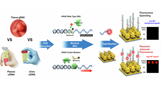
- Blood and Urine Liquid Biopsy Detects Early Colorectal Cancer Mutations
- Epigenetic Profiling Could Refine Prognosis in Acute Myeloid Leukemia
- Genetic Risk Score Supports Diagnosis and Prognosis in Idiopathic Pulmonary Fibrosis
- Extracellular Vesicle Marker Identifies Early Lung Adenocarcinoma and Predicts Recurrence
- AI Decision Support System Guides Treatment Selection for Complex Blood Cancers
- Blood Test Helps Predict Short-Term Mortality After Severe Heart Attack
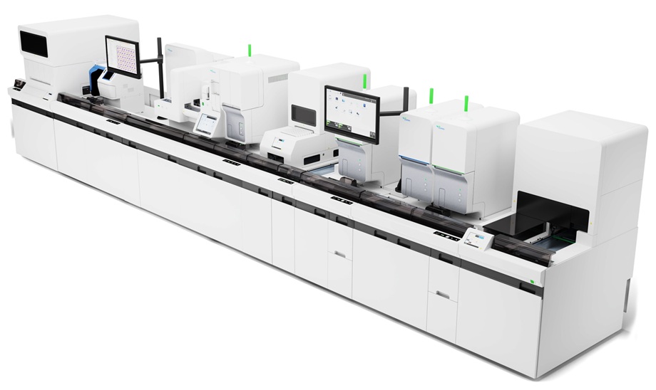
- Next-Generation Hematology Platform Streamlines High-Complexity Lab Workflows
- Blood Eosinophil Count May Predict Cancer Immunotherapy Response and Toxicity
- Higher Ferritin Threshold May Improve Iron Deficiency Detection in Children
- Anti-Lipid Antibody Biomarkers May Identify Early Lyme Disease and Persistent Symptoms
- Emergency Department Opt-Out Testing Program Identifies Undiagnosed HIV
- Immune Biomarkers Could Identify Risk of Chronic Critical Illness on ICU Admission
- New Cellular Biomarkers Correlate with Disease Severity in Sjögren Disease
- Airway Immune Signature May Predict Tuberculosis Progression Risk
- Gut Microbiome Analysis Identifies Frailty-Related Signatures in Older Adults
- CE-Marked Blood Assay Automates Tuberculosis Infection Testing
- Genomic Surveillance Algorithm Improves Early Detection of Emerging Variants
- Rapid Gastrointestinal PCR Panels Deliver One-Hour Results
- H. pylori Screening Within Colorectal Program Aids Gastric Cancer Prevention
- New CE-Certified Software Advances Whole-Genome Cancer Testing
- National Rare Disease Registry Standardizes Genetic and Clinical Data for Coordinated Care
- AI Platform Links Biomarker Results to Cancer Clinical Trials and Guidelines
- Agentic AI Platform Supports Genomic Decision-Making in Oncology
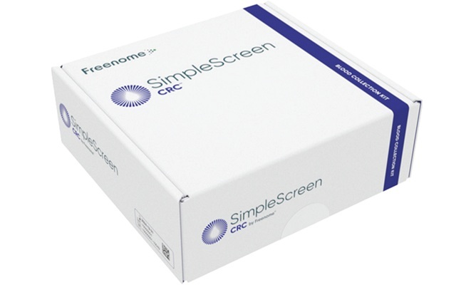
- Mailed Screening Kits Help Reduce Colorectal Cancer Screening Gaps
- Eurobio Scientific Completes Acquisition of CareDx Lab Products Division
- Blood-Based CRISPR Test for Tuberculosis Gains Regulatory Approval in Colombia
- Partnership Integrates Automated DNA Extraction with Single-Molecule Digital PCR
- Agilent Strengthens Pathology Capabilities Through Biocare Acquisition
- Merck KGaA to Acquire Bio-Techne, Expanding Life Science Portfolio
- Innovative New Technology to Provide Plastic-Exterior Components with Glass Interior, Presented at AACC 2015
- Portable Molecular Diagnostics System Unveiled At 2015 AACC
- Expanded Steroid Control Launched at the 2015 AACC Annual Meeting
- Eco-Friendly Immunoassay Reagents Featured at AACC 2015
- Low Cost Point-of-Care DNA Amplification Test for Chlamydia Infection Demonstrated at the 2015 AACC Annual Meeting
- Derived Exosomal Protein Biomarkers in Alzheimer’s Disease Diagnosis
- New Biochip Array Developed for ApoE4 Classification
- Cell-Free DNA Identifies Liver Transplant Patients with Acute Rejection
- New Method Tested for Early Diagnosis Pediatric Diabetic Nephropathy
- FDA-Cleared Automated Cell Counter for CSF Launched at AACC 2016
- Mesa Biotech Showcases PCR Testing Platform at Clinical Expo
- Abbott Diagnostics Displays Alinity Family of Systems
- Roche Diagnostics Highlights Latest Clinical Lab Solutions at AACC
- Sysmex Highlights Innovations in Flow Cytometry, Urinalysis and Hematology
- Ortho Clinical Diagnostics Unveils Next-Gen Analyzer at Lab Show
- Study Identifies Hereditary Subtype of Aggressive Prostate Cancer
- Gene Variants Linked to Pollution-Exacerbated Asthma
- Single-Cell Analysis Mapping Links Inflammation Response to Acute Myeloid Leukemia
- Study Reveals New Insights into Rare Blood Cancer Development
- New Findings Clarify Molecular Drivers of Rare Small Intestinal Cancer
- All Virtual 2020 AACC Uses Artificial Intelligence to Connect Exhibitors and Attendees
- Greiner Bio-One Showcases VACUETTE Virus Stabilization Tube for Transport and Storage of COVID-19 Swab Samples
- Stago Diagnostics Transforms Hemostasis with Innovations at AACC 2020 Virtual Event
- Roche Announces Newest Additions to Cobas Family of Analyzers at AACC 2020 Virtual Event
- Seegene Highlights High-Throughput, Single Tube 8-Plex RT-PCR Test for Flu A, Flu B, RSV and COVID-19 with Dual Internal Controls
- New Test that Measures Antibodies in Girls Who Have Received HPV Vaccine Presented at AACC 2021
- Novel Insights on COVID-19 Vaccines and Virus Evolution, AI in the Clinic, and Miniaturization of Diagnostic Platforms Explored at AACC 2021
- Seegene Unveils New STARlet-AIOS All-in-One Solution for All Molecular Testing at AACC 2021
- Group K Diagnostics Demonstrates KromaHealth Kit that Enables Lab-Quality Results at Point-of-Care
- Fluxergy Introduces First-of-Its-Kind Multi-Modal Laboratory Platform That Diagnoses COVID-19 On-Site in 60 Minutes
- Faster Method Diagnoses Pediatric Urinary Tract Infections
- Tianlong Showcases Integrated PCR Lab Solutions at AACC 2022
- Cellavision Introduces New Workflow Solution for Low-Volume Hematology Labs at AACC 2022
- Advanced Instruments Introduces New Automated Osmometer at AACC 2022
- Innovative Smartphone and AI-Based Tests Featured at AACC 2022
- Ground-Breaking Phage-Based Diagnostic Kit for Laboratory Tuberculosis Testing Presented at AACC 2023
- Laboratory Experts Show How They Are Leading the Way on Global Trends
- Unique Competition Focuses on Using Data Science to Forecast Preanalytical Errors
- Best Approach to Infectious Disease Serology Testing for Laboratorians and Clinicians Discussed at AACC 2023
- Breaking Research Throws Light on COVID, Flu, and RSV Co-Infections
- Cytovale Demonstrates 8-Minute Sepsis Diagnostic Test
- EKF Diagnostics Showcases Range of Diabetes and Hematology POC Devices
- Inpeco Launches Next-Generation Total Lab Automation System
- Tianlong Showcases Latest Products in Molecular Diagnostics, Biochemistry, Immunology and POC Testing
- Messe Düsseldorf Promotes International “MEDICAlliance” Portfolio at ADLM 2024
- Breaking Research to Make Testing for Gynecologic Cancers More Equitable and Accurate
- Clinical Data on New Assay Panel Demonstrates Accurate Assessment of Pancreatic Cancer Risk
- AI-Powered Lyme Disease Diagnostic Test Debuts at ADLM 2025
- New Study Reveals Rapid Sepsis Test Drives 56% Boost in ED Discharges
- First-Ever Fingerstick Host-Response Test Rapidly Differentiates Between Bacterial and Viral Infections
- Tissue-Based Gene Signature Signals Colorectal Cancer Recurrence Risk
- FDA-Approved Companion Diagnostic Detects PTEN Loss in Prostate Cancer
- New AI Test Delivers Rapid Breast Cancer Recurrence Predictions
- EBV Status Helps Predict Survival in Primary CNS Lymphoma
- AI Pathology Tool Predicts Immunotherapy Response in Rare Cancers







































 Client Login