





Expo
view channel
view channel
view channel
view channel
view channel
view channel
view channel
view channel
view channel
Clinical Chem.
HematologyImmunologyMicrobiologyPathologyTechnologyIndustry
Events
Webinars

- New One-Step Syphilis Test Could Enable Earlier Detection and Faster Treatment
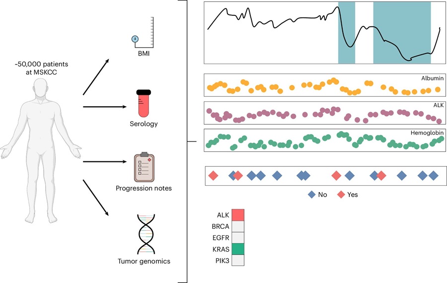
- Multimodal Clinical Data Models Predict Cachexia Risk Across Cancers
- Study Evaluates Alzheimer’s Blood Biomarker Across Racial and Ethnic Groups
- Blood Protein May Help Identify Treatment-Resistant Psychosis
- Electrochemical Biosensor Rapidly Detects Cerebrospinal Fluid Leaks
- Clinical Trial Evaluates Liquid Biopsy for Early-Stage Breast Cancer Detection
- RNA-Based Test Predicts Immunotherapy Response in Non-Small Cell Lung Cancer
- Modeling Study Shows Blood-Based CRC Screening Could Prevent More Deaths Than Stool Tests
- AI Tool Classifies Pathogenic Variants in Severe Childhood Epilepsy
- Noninvasive Cervical Test Detects Endometrial Cancer for Triage
- Handheld Point-of-Care Test Aims to Enable Rapid Coagulation Assessment in Trauma
- New Donor Genetic Marker May Help Predict Stem Cell Transplant Success
- Updated Ferritin Thresholds Improve Detection of Iron Deficiency
- New Automated Hemostasis Systems Become Available Across US Laboratories
- Variation in AML Residual Disease Testing Prompts Push for Standardization
- Immune Profiling Identifies Early Maternal Autoantibodies Linked to Preterm Birth Risk
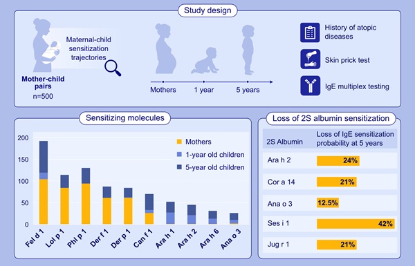
- Longitudinal Blood Testing Reveals Changes in Allergen Sensitization During Childhood
- FDA Approval Adds Automated Testing Option for Parvovirus B19 Serology
- Blood-Based Immune Aging Map Could Help Spot Disease Risk Earlier
- Blood-Based Immune Signal Could Reveal Lung Cancer Risk Before Tumors Develop
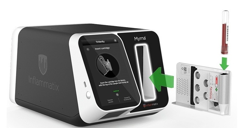
- Rapid Host-Response Test Aims to Predict Corticosteroid Benefit in Sepsis
- Oral and Lung Microbiome Changes Linked to Severe Long Covid Symptoms
- Study Finds Distinct Malaria Parasites Linked to Sickle Cell Trait Protection
- Rapid Urine Test Aids Diagnosis of Invasive Aspergillosis
- Nanodroplet CRISPR Technology Supports Rapid, Multiplexed Mycobacterial Identification
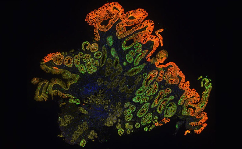
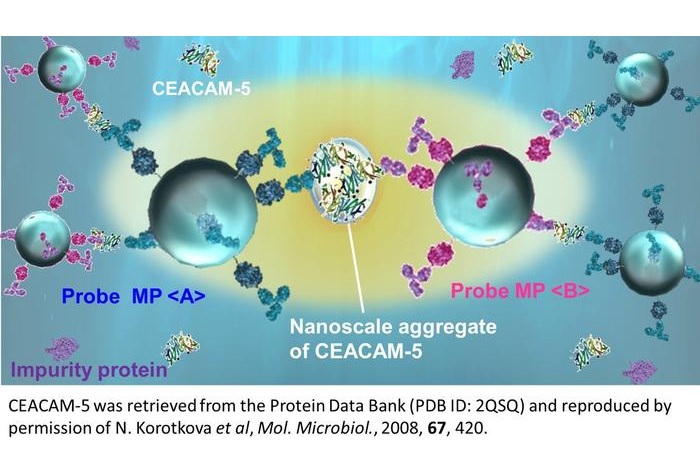
- Laser-Enhanced Assay Boosts Sensitivity for Colorectal Cancer Biomarker Detection
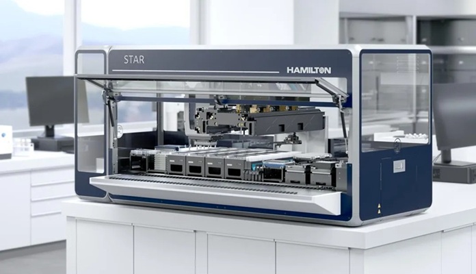
- New Automation Workflow Streamlines NGS Library Preparation for High-Volume Labs
- ADLM Calls for CLIA Updates to Support Safe AI Use in Laboratory Medicine
- Multimodal AI Framework Aims to Guide Cancer Immunotherapy Decisions
- New Multipurpose Centrifuge Combines High Capacity with Sustainable Cooling
- Myriad Genetics to Sell Prenatal Testing Portfolio to Labcorp
- Managed Care Report Examines Barriers to Breast Cancer Biomarker Testing
- Expanding Screening and Biomarker Testing Drive Cancer Diagnostics Market Growth
- Collaboration Advances Genomic Instability Biomarker for Prostate Cancer
- Labcorp Acquisition to Strengthen Specialty Testing and Prostate Cancer Assessment
- CSF Biomarker Meprin-Beta Increases in Alzheimer’s Disease
- Blood Antibody Signals May Offer Clues to the Origins of Childhood Cancer
- Cancer DNA Trapping Mechanism Reveals Potential Target for Future Therapies
- Genetic Study Links Inherited Blindness to Gene Tied to Rare Metabolic Disorder
- Population-Scale Proteomics Study Identifies New Genetic-Protein Links
- Updated Digital Pathology Platform Expands AI Integration for Cancer Diagnosis
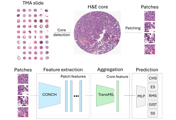
- AI Histopathology Tool Distinguishes Ewing Sarcoma for Diagnosis
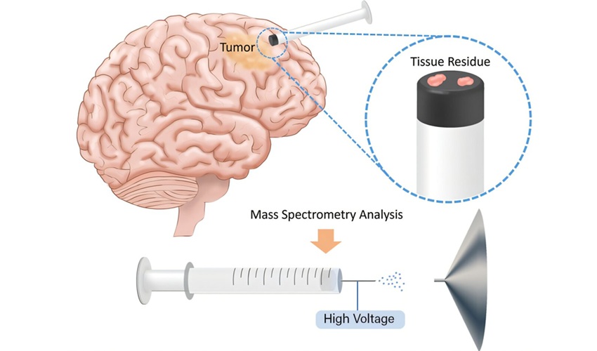
- Rapid Mass Spectrometry Test May Aid Glioma Margin Decisions
- Genomic Classifier Predicts Benefit From Adding Hormone Therapy to Salvage Prostate Radiation
- New AI Model Maps Where Glioblastoma Could Return After Surgery
 Expo
Expo
- New One-Step Syphilis Test Could Enable Earlier Detection and Faster Treatment
- Multimodal Clinical Data Models Predict Cachexia Risk Across Cancers
- Study Evaluates Alzheimer’s Blood Biomarker Across Racial and Ethnic Groups
- Blood Protein May Help Identify Treatment-Resistant Psychosis
- Electrochemical Biosensor Rapidly Detects Cerebrospinal Fluid Leaks
- Clinical Trial Evaluates Liquid Biopsy for Early-Stage Breast Cancer Detection
- RNA-Based Test Predicts Immunotherapy Response in Non-Small Cell Lung Cancer
- Modeling Study Shows Blood-Based CRC Screening Could Prevent More Deaths Than Stool Tests
- AI Tool Classifies Pathogenic Variants in Severe Childhood Epilepsy
- Noninvasive Cervical Test Detects Endometrial Cancer for Triage
- Handheld Point-of-Care Test Aims to Enable Rapid Coagulation Assessment in Trauma
- New Donor Genetic Marker May Help Predict Stem Cell Transplant Success
- Updated Ferritin Thresholds Improve Detection of Iron Deficiency
- New Automated Hemostasis Systems Become Available Across US Laboratories
- Variation in AML Residual Disease Testing Prompts Push for Standardization
- Immune Profiling Identifies Early Maternal Autoantibodies Linked to Preterm Birth Risk
- Longitudinal Blood Testing Reveals Changes in Allergen Sensitization During Childhood
- FDA Approval Adds Automated Testing Option for Parvovirus B19 Serology
- Blood-Based Immune Aging Map Could Help Spot Disease Risk Earlier
- Blood-Based Immune Signal Could Reveal Lung Cancer Risk Before Tumors Develop
- Rapid Host-Response Test Aims to Predict Corticosteroid Benefit in Sepsis
- Oral and Lung Microbiome Changes Linked to Severe Long Covid Symptoms
- Study Finds Distinct Malaria Parasites Linked to Sickle Cell Trait Protection
- Rapid Urine Test Aids Diagnosis of Invasive Aspergillosis
- Nanodroplet CRISPR Technology Supports Rapid, Multiplexed Mycobacterial Identification
- Laser-Enhanced Assay Boosts Sensitivity for Colorectal Cancer Biomarker Detection
- New Automation Workflow Streamlines NGS Library Preparation for High-Volume Labs
- ADLM Calls for CLIA Updates to Support Safe AI Use in Laboratory Medicine
- Multimodal AI Framework Aims to Guide Cancer Immunotherapy Decisions
- New Multipurpose Centrifuge Combines High Capacity with Sustainable Cooling
- Myriad Genetics to Sell Prenatal Testing Portfolio to Labcorp
- Managed Care Report Examines Barriers to Breast Cancer Biomarker Testing
- Expanding Screening and Biomarker Testing Drive Cancer Diagnostics Market Growth
- Collaboration Advances Genomic Instability Biomarker for Prostate Cancer
- Labcorp Acquisition to Strengthen Specialty Testing and Prostate Cancer Assessment
- CSF Biomarker Meprin-Beta Increases in Alzheimer’s Disease
- Blood Antibody Signals May Offer Clues to the Origins of Childhood Cancer
- Cancer DNA Trapping Mechanism Reveals Potential Target for Future Therapies
- Genetic Study Links Inherited Blindness to Gene Tied to Rare Metabolic Disorder
- Population-Scale Proteomics Study Identifies New Genetic-Protein Links
- Updated Digital Pathology Platform Expands AI Integration for Cancer Diagnosis
- AI Histopathology Tool Distinguishes Ewing Sarcoma for Diagnosis
- Rapid Mass Spectrometry Test May Aid Glioma Margin Decisions
- Genomic Classifier Predicts Benefit From Adding Hormone Therapy to Salvage Prostate Radiation
- New AI Model Maps Where Glioblastoma Could Return After Surgery





































 Client Login