




Expo
view channel
view channel
view channel
view channel
view channel
view channel
view channel
view channel
view channel
Clinical Chem.Molecular DiagnosticsHematologyImmunologyMicrobiologyPathology
Industry
Events
Webinars

- Blood Proteins Reveal Signals Linked to Prostate Cancer Risk Years Before Diagnosis
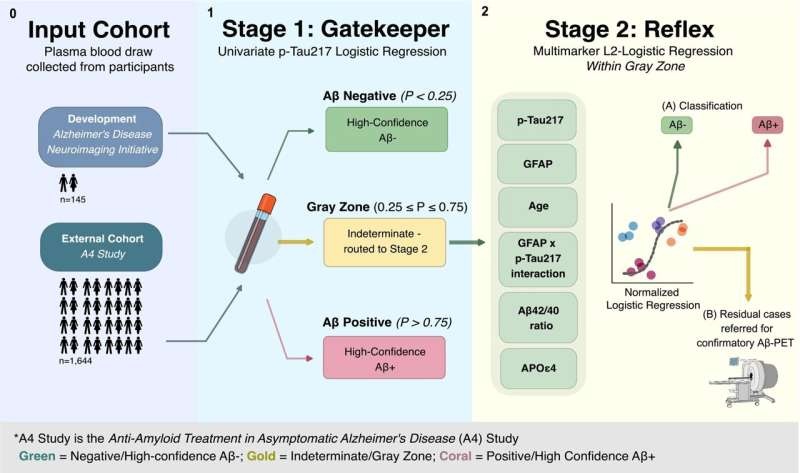
- Two-Step Blood Testing Approach Reduces Uncertainty in Amyloid Assessment
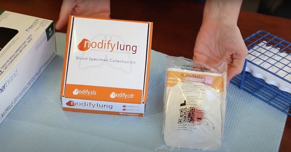
- Multimodal Platform to Combine Blood and Imaging Biomarkers for Lung Nodule Assessment
- Study Links Saliva-Based Epigenetic Signals to OCD Treatment Outcomes
- New One-Step Syphilis Test Could Enable Earlier Detection and Faster Treatment
- New Variant Tracking Option Expands Molecular Residual Disease Monitoring
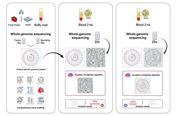
- Liquid Biopsy Detects Lung Cancer Recurrence Signals Months Before Imaging
- GeneDx Presents Evidence for Wider Clinical Use of Genomic Sequencing at ASHG 2026
- Streamlined Genetic Testing Speeds Pediatric Immune Disorder Diagnosis
- Clinical Trial Evaluates Liquid Biopsy for Early-Stage Breast Cancer Detection
- Handheld Point-of-Care Test Aims to Enable Rapid Coagulation Assessment in Trauma
- New Donor Genetic Marker May Help Predict Stem Cell Transplant Success
- Updated Ferritin Thresholds Improve Detection of Iron Deficiency
- New Automated Hemostasis Systems Become Available Across US Laboratories
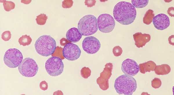
- Variation in AML Residual Disease Testing Prompts Push for Standardization
- Immune Profiling Initiative Aims to Improve Bone Marrow Analysis in Leukemia
- Immune Profiling Identifies Early Maternal Autoantibodies Linked to Preterm Birth Risk
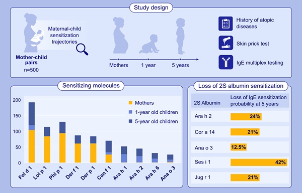
- Longitudinal Blood Testing Reveals Changes in Allergen Sensitization During Childhood
- FDA Approval Adds Automated Testing Option for Parvovirus B19 Serology
- Blood-Based Immune Aging Map Could Help Spot Disease Risk Earlier
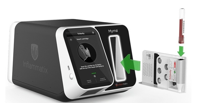
- Rapid Host-Response Test Aims to Predict Corticosteroid Benefit in Sepsis
- Oral and Lung Microbiome Changes Linked to Severe Long Covid Symptoms
- Study Finds Distinct Malaria Parasites Linked to Sickle Cell Trait Protection
- Rapid Urine Test Aids Diagnosis of Invasive Aspergillosis
- Nanodroplet CRISPR Technology Supports Rapid, Multiplexed Mycobacterial Identification
- Laser-Based Swab Analysis Shows Promise for Detecting Disease-Linked Odor Patterns
- Laser-Enhanced Assay Boosts Sensitivity for Colorectal Cancer Biomarker Detection
- New Automation Workflow Streamlines NGS Library Preparation for High-Volume Labs
- ADLM Calls for CLIA Updates to Support Safe AI Use in Laboratory Medicine
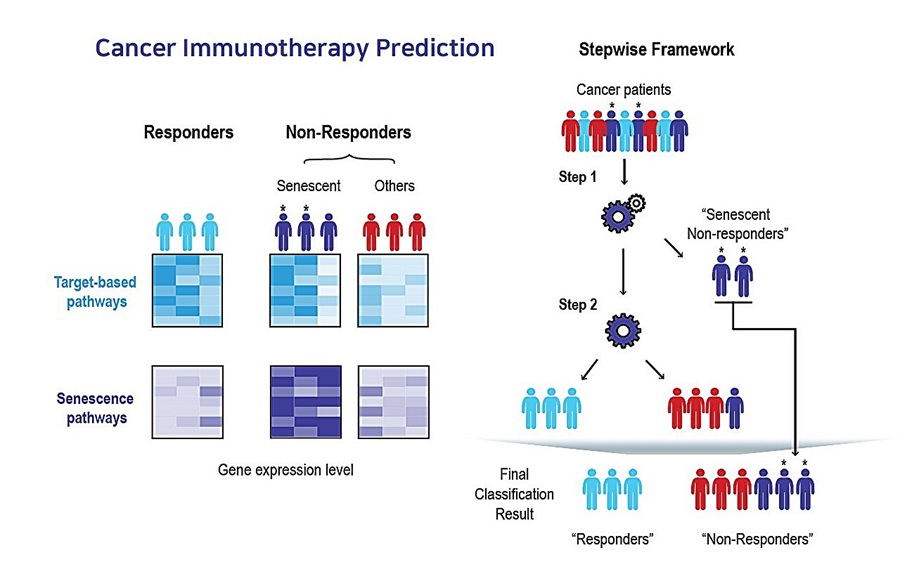
- Multimodal AI Framework Aims to Guide Cancer Immunotherapy Decisions
- Alzheimer’s Progression Blood Test Gains U.S. Patent Ahead of Planned Launch
- Hologic to Broaden Breast Cancer Testing Portfolio Through Reveal Genomics Acquisition
- Myriad Genetics to Sell Prenatal Testing Portfolio to Labcorp
- Managed Care Report Examines Barriers to Breast Cancer Biomarker Testing
- Expanding Screening and Biomarker Testing Drive Cancer Diagnostics Market Growth
- Genomic Tool Supports Analysis of Repeat Expansions Linked to Inherited Disorders
- CSF Biomarker Meprin-Beta Increases in Alzheimer’s Disease
- Blood Antibody Signals May Offer Clues to the Origins of Childhood Cancer
- Cancer DNA Trapping Mechanism Reveals Potential Target for Future Therapies
- Genetic Study Links Inherited Blindness to Gene Tied to Rare Metabolic Disorder
- AI Pre-Screening Helps Identify Lung Cancer Cases for Additional Biomarker Testing
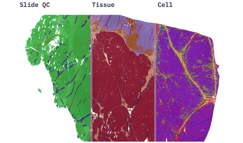
- AI Pathology Platform Targets Workflow Delays and Molecular Test Failures
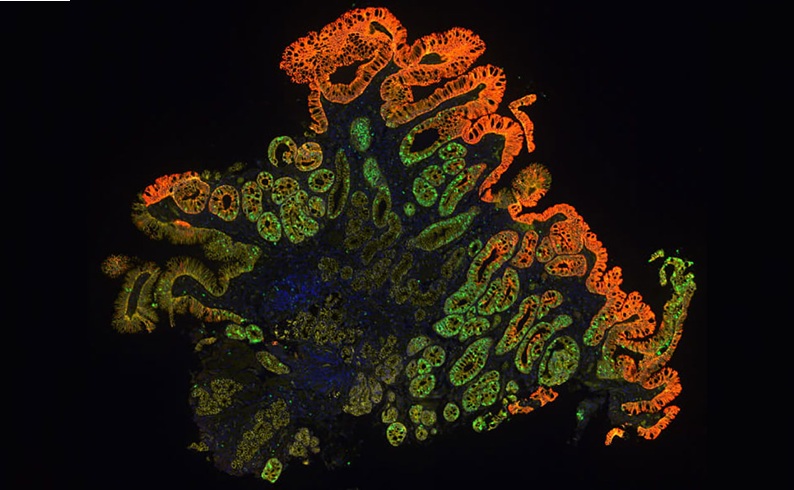
- Molecular Testing Refines Barrett’s Esophagus Progression Risk Beyond Biopsy Findings
- Updated Digital Pathology Platform Expands AI Integration for Cancer Diagnosis
- AI Histopathology Tool Distinguishes Ewing Sarcoma for Diagnosis
 Expo
Expo
- Blood Proteins Reveal Signals Linked to Prostate Cancer Risk Years Before Diagnosis
- Two-Step Blood Testing Approach Reduces Uncertainty in Amyloid Assessment
- Multimodal Platform to Combine Blood and Imaging Biomarkers for Lung Nodule Assessment
- Study Links Saliva-Based Epigenetic Signals to OCD Treatment Outcomes
- New One-Step Syphilis Test Could Enable Earlier Detection and Faster Treatment
- New Variant Tracking Option Expands Molecular Residual Disease Monitoring
- Liquid Biopsy Detects Lung Cancer Recurrence Signals Months Before Imaging
- GeneDx Presents Evidence for Wider Clinical Use of Genomic Sequencing at ASHG 2026
- Streamlined Genetic Testing Speeds Pediatric Immune Disorder Diagnosis
- Clinical Trial Evaluates Liquid Biopsy for Early-Stage Breast Cancer Detection
- Handheld Point-of-Care Test Aims to Enable Rapid Coagulation Assessment in Trauma
- New Donor Genetic Marker May Help Predict Stem Cell Transplant Success
- Updated Ferritin Thresholds Improve Detection of Iron Deficiency
- New Automated Hemostasis Systems Become Available Across US Laboratories
- Variation in AML Residual Disease Testing Prompts Push for Standardization
- Immune Profiling Initiative Aims to Improve Bone Marrow Analysis in Leukemia
- Immune Profiling Identifies Early Maternal Autoantibodies Linked to Preterm Birth Risk
- Longitudinal Blood Testing Reveals Changes in Allergen Sensitization During Childhood
- FDA Approval Adds Automated Testing Option for Parvovirus B19 Serology
- Blood-Based Immune Aging Map Could Help Spot Disease Risk Earlier
- Rapid Host-Response Test Aims to Predict Corticosteroid Benefit in Sepsis
- Oral and Lung Microbiome Changes Linked to Severe Long Covid Symptoms
- Study Finds Distinct Malaria Parasites Linked to Sickle Cell Trait Protection
- Rapid Urine Test Aids Diagnosis of Invasive Aspergillosis
- Nanodroplet CRISPR Technology Supports Rapid, Multiplexed Mycobacterial Identification
- Laser-Based Swab Analysis Shows Promise for Detecting Disease-Linked Odor Patterns
- Laser-Enhanced Assay Boosts Sensitivity for Colorectal Cancer Biomarker Detection
- New Automation Workflow Streamlines NGS Library Preparation for High-Volume Labs
- ADLM Calls for CLIA Updates to Support Safe AI Use in Laboratory Medicine
- Multimodal AI Framework Aims to Guide Cancer Immunotherapy Decisions
- Alzheimer’s Progression Blood Test Gains U.S. Patent Ahead of Planned Launch
- Hologic to Broaden Breast Cancer Testing Portfolio Through Reveal Genomics Acquisition
- Myriad Genetics to Sell Prenatal Testing Portfolio to Labcorp
- Managed Care Report Examines Barriers to Breast Cancer Biomarker Testing
- Expanding Screening and Biomarker Testing Drive Cancer Diagnostics Market Growth
- Genomic Tool Supports Analysis of Repeat Expansions Linked to Inherited Disorders
- CSF Biomarker Meprin-Beta Increases in Alzheimer’s Disease
- Blood Antibody Signals May Offer Clues to the Origins of Childhood Cancer
- Cancer DNA Trapping Mechanism Reveals Potential Target for Future Therapies
- Genetic Study Links Inherited Blindness to Gene Tied to Rare Metabolic Disorder
- AI Pre-Screening Helps Identify Lung Cancer Cases for Additional Biomarker Testing
- AI Pathology Platform Targets Workflow Delays and Molecular Test Failures
- Molecular Testing Refines Barrett’s Esophagus Progression Risk Beyond Biopsy Findings
- Updated Digital Pathology Platform Expands AI Integration for Cancer Diagnosis
- AI Histopathology Tool Distinguishes Ewing Sarcoma for Diagnosis








































 Client Login