




Expo
view channel
view channel
view channel
view channel
view channel
view channel
view channel
view channel
view channel
Clinical Chem.Molecular DiagnosticsHematologyImmunologyMicrobiology
TechnologyIndustry
Events
Webinars

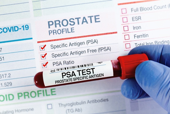
- Updated PSA Testing Guidelines Support Early Prostate Cancer Detection
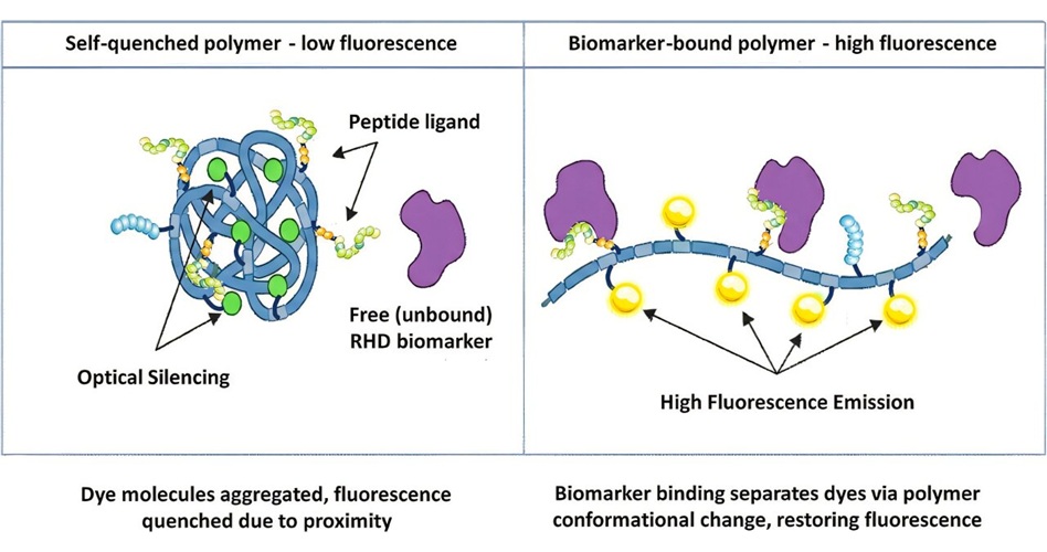
- Antibody-Free Blood Test Detects Rheumatic Heart Disease Biomarker
- Blood and Spinal Fluid Biomarker May Help Track Early Huntington’s Disease
- New Biomarker Offers Longer Detection Window for Suspected GHB Exposure
- Study Finds Urine Biomarker Score Improves Five-Year Cancer Risk Prediction
- AI in Cancer Diagnosis and Molecular Testing Takes Center Stage at AMP 2026
- AMP 2026 Highlights Advances in Molecular Testing for Pediatric Diseases
- Genomic Studies Investigate Treatment Response and Inherited Risk in Sarcoma
- Liquid Biopsy Expands Insights Into Treatment Response and Tumor Alterations
- Genomic Sequencing Identifies Actionable Findings to Guide Rare Disease Management
- Study Confirms Blood Products Remain Viable After Three Weeks in the Arctic
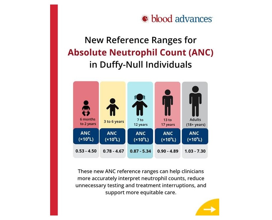
- New Reference Ranges Clarify Normal Neutrophil Counts in Duffy-Null Patients
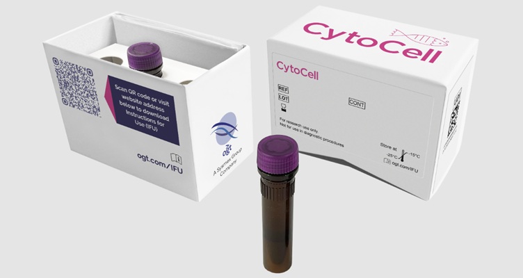
- Newly Certified FISH Probes Support Cytogenetic Testing in Blood Cancers
- Genetic Testing Offers Opportunities for Earlier Blood Disorder Carrier Identification
- New Risk-Stratification Tool Predicts Relapse After Blinatumomab in Childhood ALL
- Antibody Testing Informs Melioidosis Diagnosis and Surveillance in Emerging Regions
- Exhaled Breath Particles Reveal Virus-Specific Antibodies in the Lower Airways
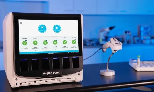
- Roche Gains FDA Clearance for Flexible Respiratory Pathogen Panel
- Tumor and Immune Biomarkers Linked to Treatment Response in T-Cell Lymphoma
- Prototype Point-of-Care Lateral Flow Test Aims to Improve Neonatal Sepsis Triage
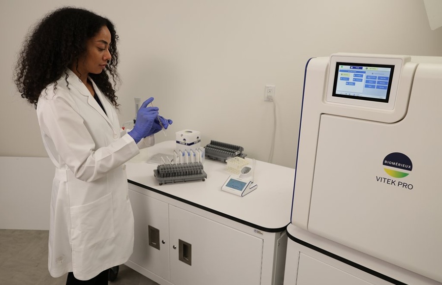
- Automated Microbial Identification and Susceptibility System Gains FDA Clearance
- Gastrointestinal Flex Assay Gains EU Certification for Tailored Pathogen Testing
- Molecular Panels Gain FDA Clearance for One-Hour Gastrointestinal Pathogen Testing
- Common Genetic Variant Increases Risk of Bacterial Pneumonia
- Stool Testing Predicts UTI Risk and Guides Antibiotic Selection
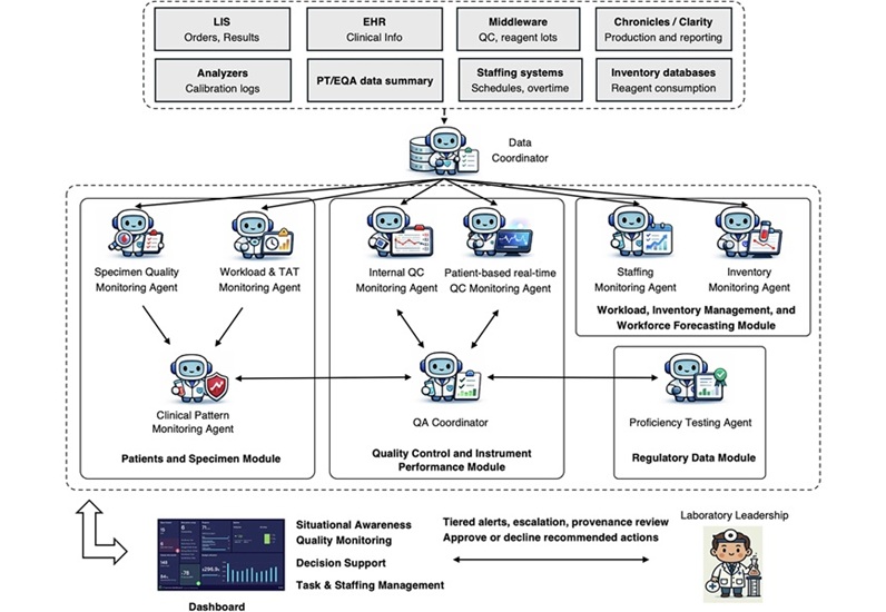
- Multi-Agent AI Framework Supports Laboratory Quality and Workflow Monitoring
- Personal PCR Analyzer Aims to Enable At-Home Infectious Disease Testing
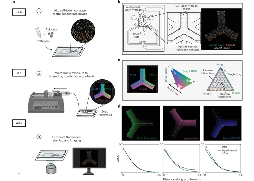
- Microfluidic Assay Predicts Treatment Response in Pediatric Leukemia
- AI Models Use Routine Blood Tests and Health Records to Predict Cancer Risk
- Compact PCR Thermal Cycler Launches for Routine Molecular Laboratory Workflows
- Partnership Aims to Bring Blood-Based Alzheimer’s Testing to Japan
- Siemens Healthineers Begins Transition Toward Standalone Olyrian Diagnostics Business
- Agilent Completes Acquisition of Digital Pathology Platform Developer Proscia
- New Diagnostics Company Onvaro Formed to Expand Decentralized Testing
- Illumina Selected to Support National Genomics Study for Personalized Healthcare in Singapore
- Extracellular Vesicle Biomarkers Reveal Shared Signals in Aging and Cancer
- Multiplexed Single-Cell DNA Methylation Workflow Enables Analysis of Blood Disorders
- Study Reveals Regulatory DNA Element Involved in Breast Cancer Growth Suppression
- Platelet Biomarkers Identify Progression Signals in Parkinson's Disease
- New ddPCR Assays Broaden Options for Multiplex Cancer Mutation Research
- Review Highlights Progress and Validation Needs for AI in Digital Pathology
- Breakthrough-Designated Cytology Test Supports Oral Cancer Risk Assessment
- AI Pathology Approach Closely Aligns With Breast Cancer Genomic Risk Classification
- Roche Launches Automated Test to Support Mesothelioma Differential Diagnosis
- Digital Pathology Test Stratifies Recurrence Risk in Early Breast Cancer
 Expo
- Updated PSA Testing Guidelines Support Early Prostate Cancer Detection
- Antibody-Free Blood Test Detects Rheumatic Heart Disease Biomarker
- Blood and Spinal Fluid Biomarker May Help Track Early Huntington’s Disease
- New Biomarker Offers Longer Detection Window for Suspected GHB Exposure
- Study Finds Urine Biomarker Score Improves Five-Year Cancer Risk Prediction
- AI in Cancer Diagnosis and Molecular Testing Takes Center Stage at AMP 2026
- AMP 2026 Highlights Advances in Molecular Testing for Pediatric Diseases
- Genomic Studies Investigate Treatment Response and Inherited Risk in Sarcoma
- Liquid Biopsy Expands Insights Into Treatment Response and Tumor Alterations
- Genomic Sequencing Identifies Actionable Findings to Guide Rare Disease Management
- Study Confirms Blood Products Remain Viable After Three Weeks in the Arctic
- New Reference Ranges Clarify Normal Neutrophil Counts in Duffy-Null Patients
- Newly Certified FISH Probes Support Cytogenetic Testing in Blood Cancers
- Genetic Testing Offers Opportunities for Earlier Blood Disorder Carrier Identification
- New Risk-Stratification Tool Predicts Relapse After Blinatumomab in Childhood ALL
- Antibody Testing Informs Melioidosis Diagnosis and Surveillance in Emerging Regions
- Exhaled Breath Particles Reveal Virus-Specific Antibodies in the Lower Airways
- Roche Gains FDA Clearance for Flexible Respiratory Pathogen Panel
- Tumor and Immune Biomarkers Linked to Treatment Response in T-Cell Lymphoma
- Prototype Point-of-Care Lateral Flow Test Aims to Improve Neonatal Sepsis Triage
- Automated Microbial Identification and Susceptibility System Gains FDA Clearance
- Gastrointestinal Flex Assay Gains EU Certification for Tailored Pathogen Testing
- Molecular Panels Gain FDA Clearance for One-Hour Gastrointestinal Pathogen Testing
- Common Genetic Variant Increases Risk of Bacterial Pneumonia
- Stool Testing Predicts UTI Risk and Guides Antibiotic Selection
- Multi-Agent AI Framework Supports Laboratory Quality and Workflow Monitoring
- Personal PCR Analyzer Aims to Enable At-Home Infectious Disease Testing
- Microfluidic Assay Predicts Treatment Response in Pediatric Leukemia
- AI Models Use Routine Blood Tests and Health Records to Predict Cancer Risk
- Compact PCR Thermal Cycler Launches for Routine Molecular Laboratory Workflows
- Partnership Aims to Bring Blood-Based Alzheimer’s Testing to Japan
- Siemens Healthineers Begins Transition Toward Standalone Olyrian Diagnostics Business
- Agilent Completes Acquisition of Digital Pathology Platform Developer Proscia
- New Diagnostics Company Onvaro Formed to Expand Decentralized Testing
- Illumina Selected to Support National Genomics Study for Personalized Healthcare in Singapore
- Extracellular Vesicle Biomarkers Reveal Shared Signals in Aging and Cancer
- Multiplexed Single-Cell DNA Methylation Workflow Enables Analysis of Blood Disorders
- Study Reveals Regulatory DNA Element Involved in Breast Cancer Growth Suppression
- Platelet Biomarkers Identify Progression Signals in Parkinson's Disease
- New ddPCR Assays Broaden Options for Multiplex Cancer Mutation Research
- Review Highlights Progress and Validation Needs for AI in Digital Pathology
- Breakthrough-Designated Cytology Test Supports Oral Cancer Risk Assessment
- AI Pathology Approach Closely Aligns With Breast Cancer Genomic Risk Classification
- Roche Launches Automated Test to Support Mesothelioma Differential Diagnosis
- Digital Pathology Test Stratifies Recurrence Risk in Early Breast Cancer






























 Client Login