




Expo
view channel
view channel
view channel
view channel
view channel
view channel
view channel
view channel
view channel
Clinical Chem.Molecular DiagnosticsHematologyImmunologyMicrobiologyPathology
Industry
Events
Webinars

- Plasma Proteomic Test Detects Immunotherapy Resistance Before Imaging in Lung Cancer
- Blood Protein Analysis Reveals Menopause-Associated Signals of Brain Aging
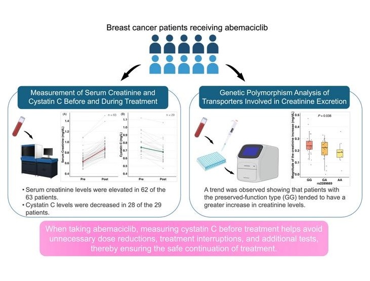
- Cystatin C Testing Helps Distinguish Abemaciclib Effect from Kidney Injury
- Blood-Based Score May Support Targeted Interventions for Healthier Aging
- Noninvasive Tests Offer Alternatives to Biopsy for Assessing Advanced Liver Scarring
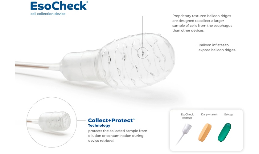
- Nonendoscopic Methylation Assay Enables Rapid Barrett’s Esophagus Screening
- Studies Support MRD Testing to Detect Head and Neck Cancer Recurrence
- Genomic Profiling Predicts Relapse Risk by Subtype in Pediatric Leukemia
- AI-Supported Approach May Improve Cervical Cancer Screening in Low-Resource Settings
- Molecular AI Model Refines Risk Prediction in Pediatric Brain Tumors
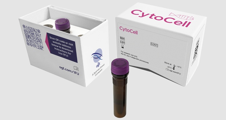
- Newly Certified FISH Probes Support Cytogenetic Testing in Blood Cancers
- Genetic Testing Offers Opportunities for Earlier Blood Disorder Carrier Identification
- New Risk-Stratification Tool Predicts Relapse After Blinatumomab in Childhood ALL
- New Myelofibrosis Guidelines Recommend Expanded Genetic Testing at Diagnosis
- Handheld Point-of-Care Test Aims to Enable Rapid Coagulation Assessment in Trauma
- Roche Gains FDA Clearance for Flexible Respiratory Pathogen Panel
- Tumor and Immune Biomarkers Linked to Treatment Response in T-Cell Lymphoma
- Prototype Point-of-Care Lateral Flow Test Aims to Improve Neonatal Sepsis Triage
- Immune and Inflammatory Markers May Help Assess CAR T-Cell Complication Risk
- COVID-19 and Flu Combo Test Launches With FDA Clearance
- Rapid Host-Response Assay Gains FDA Clearance to Help Distinguish Bacterial and Viral Infections
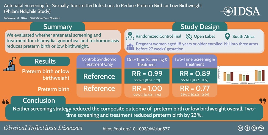
- Repeat Prenatal STI Screening and Treatment May Reduce Preterm Births
- Decentralized Laboratory Network Improves Detection of Viral Hemorrhagic Fevers in Guinea
- Molecular Panel Gains WHO Emergency Use Listing to Support Ebola Outbreak Response
- Rapid Host-Response Test Aims to Predict Corticosteroid Benefit in Sepsis
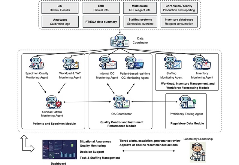
- Multi-Agent AI Framework Supports Laboratory Quality and Workflow Monitoring
- Personal PCR Analyzer Aims to Enable At-Home Infectious Disease Testing
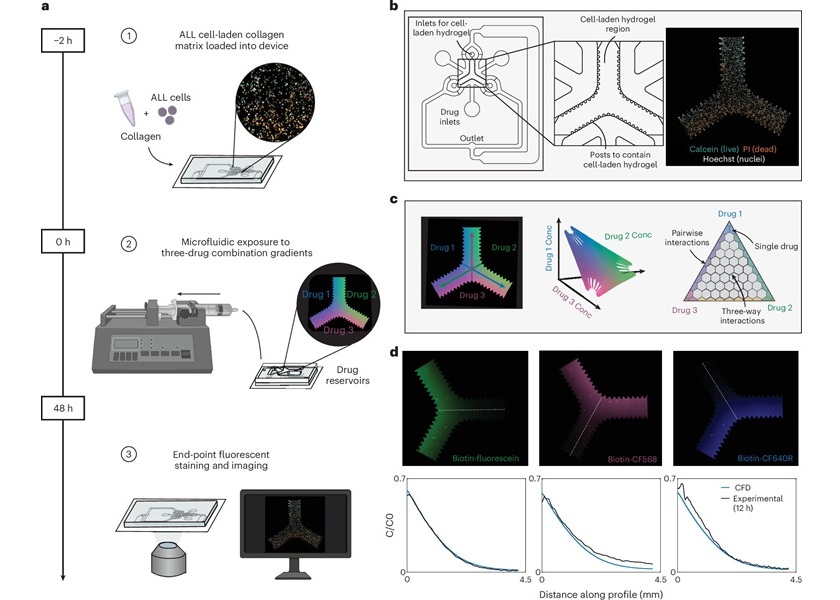
- Microfluidic Assay Predicts Treatment Response in Pediatric Leukemia
- AI Models Use Routine Blood Tests and Health Records to Predict Cancer Risk
- Compact PCR Thermal Cycler Launches for Routine Molecular Laboratory Workflows
- Agilent Completes Acquisition of Digital Pathology Platform Developer Proscia
- New Diagnostics Company Onvaro Formed to Expand Decentralized Testing
- Illumina Selected to Support National Genomics Study for Personalized Healthcare in Singapore
- Collaboration Expands Access to Tests for Cancer Treatment Resistance and Toxicity Risk
- CE-Marked IVD Sequencing Platform Launches Across Ten EU Countries
- New ddPCR Assays Broaden Options for Multiplex Cancer Mutation Research
- Molecular Atlas Offers New Framework for Breast Cancer Risk and Treatment Research
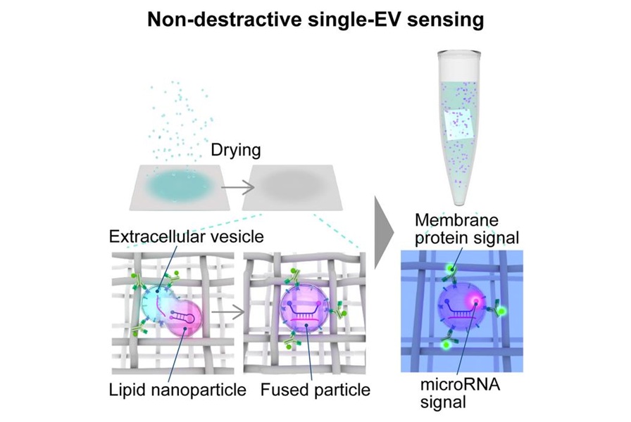
- New Method Isolates Neuron-Linked Vesicles From Blood and Cerebrospinal Fluid
- Lung Tumor RNA Variants Offer Clues to Immunotherapy Benefit
- Population-Scale Genome Survey Supports Interpretation of Genetic Testing
- Breakthrough-Designated Cytology Test Supports Oral Cancer Risk Assessment
- AI Pathology Approach Closely Aligns With Breast Cancer Genomic Risk Classification
- Roche Launches Automated Test to Support Mesothelioma Differential Diagnosis
- Digital Pathology Test Stratifies Recurrence Risk in Early Breast Cancer
- AI Pathology Tool Predicts DNA Repair Deficiency in High-Grade Serous Ovarian Cancer
 Expo
- Plasma Proteomic Test Detects Immunotherapy Resistance Before Imaging in Lung Cancer
- Blood Protein Analysis Reveals Menopause-Associated Signals of Brain Aging
- Cystatin C Testing Helps Distinguish Abemaciclib Effect from Kidney Injury
- Blood-Based Score May Support Targeted Interventions for Healthier Aging
- Noninvasive Tests Offer Alternatives to Biopsy for Assessing Advanced Liver Scarring
- Nonendoscopic Methylation Assay Enables Rapid Barrett’s Esophagus Screening
- Studies Support MRD Testing to Detect Head and Neck Cancer Recurrence
- Genomic Profiling Predicts Relapse Risk by Subtype in Pediatric Leukemia
- AI-Supported Approach May Improve Cervical Cancer Screening in Low-Resource Settings
- Molecular AI Model Refines Risk Prediction in Pediatric Brain Tumors
- Newly Certified FISH Probes Support Cytogenetic Testing in Blood Cancers
- Genetic Testing Offers Opportunities for Earlier Blood Disorder Carrier Identification
- New Risk-Stratification Tool Predicts Relapse After Blinatumomab in Childhood ALL
- New Myelofibrosis Guidelines Recommend Expanded Genetic Testing at Diagnosis
- Handheld Point-of-Care Test Aims to Enable Rapid Coagulation Assessment in Trauma
- Roche Gains FDA Clearance for Flexible Respiratory Pathogen Panel
- Tumor and Immune Biomarkers Linked to Treatment Response in T-Cell Lymphoma
- Prototype Point-of-Care Lateral Flow Test Aims to Improve Neonatal Sepsis Triage
- Immune and Inflammatory Markers May Help Assess CAR T-Cell Complication Risk
- COVID-19 and Flu Combo Test Launches With FDA Clearance
- Rapid Host-Response Assay Gains FDA Clearance to Help Distinguish Bacterial and Viral Infections
- Repeat Prenatal STI Screening and Treatment May Reduce Preterm Births
- Decentralized Laboratory Network Improves Detection of Viral Hemorrhagic Fevers in Guinea
- Molecular Panel Gains WHO Emergency Use Listing to Support Ebola Outbreak Response
- Rapid Host-Response Test Aims to Predict Corticosteroid Benefit in Sepsis
- Multi-Agent AI Framework Supports Laboratory Quality and Workflow Monitoring
- Personal PCR Analyzer Aims to Enable At-Home Infectious Disease Testing
- Microfluidic Assay Predicts Treatment Response in Pediatric Leukemia
- AI Models Use Routine Blood Tests and Health Records to Predict Cancer Risk
- Compact PCR Thermal Cycler Launches for Routine Molecular Laboratory Workflows
- Agilent Completes Acquisition of Digital Pathology Platform Developer Proscia
- New Diagnostics Company Onvaro Formed to Expand Decentralized Testing
- Illumina Selected to Support National Genomics Study for Personalized Healthcare in Singapore
- Collaboration Expands Access to Tests for Cancer Treatment Resistance and Toxicity Risk
- CE-Marked IVD Sequencing Platform Launches Across Ten EU Countries
- New ddPCR Assays Broaden Options for Multiplex Cancer Mutation Research
- Molecular Atlas Offers New Framework for Breast Cancer Risk and Treatment Research
- New Method Isolates Neuron-Linked Vesicles From Blood and Cerebrospinal Fluid
- Lung Tumor RNA Variants Offer Clues to Immunotherapy Benefit
- Population-Scale Genome Survey Supports Interpretation of Genetic Testing
- Breakthrough-Designated Cytology Test Supports Oral Cancer Risk Assessment
- AI Pathology Approach Closely Aligns With Breast Cancer Genomic Risk Classification
- Roche Launches Automated Test to Support Mesothelioma Differential Diagnosis
- Digital Pathology Test Stratifies Recurrence Risk in Early Breast Cancer
- AI Pathology Tool Predicts DNA Repair Deficiency in High-Grade Serous Ovarian Cancer
































 Client Login