




Expo
view channel
view channel
view channel
view channel
view channel
view channel
view channel
view channel
view channel
Clinical Chem.Molecular DiagnosticsHematologyImmunologyMicrobiology
TechnologyIndustry
Events
Webinars

- Study Questions Broad Fasting Requirements Before Routine Blood Tests
- Elevated Lipoprotein(a) Linked to Long-Term Progression of Carotid Atherosclerosis
- Lollipop-Inspired Device Measures Acetaminophen Levels in Saliva
- New Insights Into Fetal DNA Could Improve Non-Invasive Prenatal Testing
- UK Project Advances Blood-Based Dementia Testing Toward Routine Clinical Care
- Age-Based Genetic Testing May Miss Most Inherited Cancer Risk Variants
- New NGS Suite Expands Clonality and MRD Research in Lymphoid Malignancies
- Tumor-Informed ctDNA Test Shows Utility Across Gastrointestinal and Gynecologic Cancers
- Pooled Sputum Testing Offers Lower-Cost Molecular Diagnosis for Tuberculosis
- Tumor Markers Help Predict Response to Less Intensive HER2+ Chemotherapy
- Updated Ferritin Thresholds Improve Detection of Iron Deficiency
- New Automated Hemostasis Systems Become Available Across US Laboratories
- Variation in AML Residual Disease Testing Prompts Push for Standardization
- Hidden Molecular Mechanism Behind HIT Blood-Clotting Disorder Revealed
- Borderline Anemia May Carry Higher Risk Than Current Thresholds Suggest
- Prototype Point-of-Care Test Aims to Improve Neonatal Sepsis Diagnosis
- Single-Tier Lyme Disease Test Shows High Sensitivity in Early Infection
- New Web-Based Platform Helps Autoimmune Laboratories Manage Testing Complexity
- Routine Newborn Blood Spots Can Measure GBS Antibody Levels
- Blood Test Differentiates Bacterial and Viral Infections in Febrile Infants
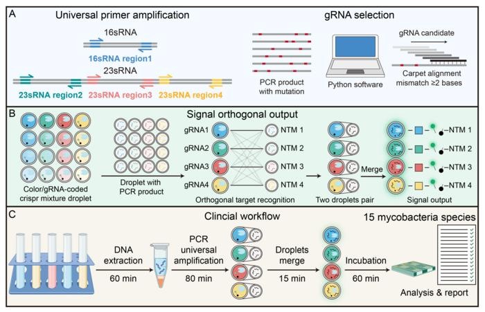
- Nanodroplet CRISPR Technology Supports Rapid, Multiplexed Mycobacterial Identification
- Genomic Workflow Identifies Fungal Pathogens Before Blood Cultures Turn Positive
- FDA-Cleared Multiplex PCR Test Detects 13 Respiratory Pathogens in a Single Sample
- One-Hour Molecular Panel Expands Bloodstream Infection Testing for Gram-Negative Pathogens
- FDA Clears Rapid Phenotypic Antimicrobial Susceptibility System for Positive Blood Cultures
- Laser-Enhanced Assay Boosts Sensitivity for Colorectal Cancer Biomarker Detection
- New Automation Workflow Streamlines NGS Library Preparation for High-Volume Labs
- ADLM Calls for CLIA Updates to Support Safe AI Use in Laboratory Medicine
- Multimodal AI Framework Aims to Guide Cancer Immunotherapy Decisions
- New Multipurpose Centrifuge Combines High Capacity with Sustainable Cooling
- Collaboration Advances Automated Benchtop Platform for Routine Blood Testing
- Expanded Partnership Supports AI Biomarker Validation and Clinical Trial Deployment
- Collaboration Combines AI Cognitive Assessment and RNA Blood Testing for Earlier Alzheimer’s Detection
- Mayo Clinic Laboratories and Pathology Asia Expand Genomic Testing Across Asia-Pacific
- New UK Collaboration Expands Access to Integrated Alzheimer’s Diagnostic Testing
- Population-Scale Proteomics Study Identifies New Genetic-Protein Links
- Circulating Tumor DNA Helps Identify Targets for Personalized Immunotherapy
- Researchers Classify 108 Lysosomal Disorders in Updated Diagnostic Framework
- New Autoantibody Target Identified in Neuromyelitis Optica Spectrum Disorder
- New Gene-Disease Link May Help Diagnose Rare Neurodevelopmental Disorders
- Rapid Mass Spectrometry Test May Aid Glioma Margin Decisions
- Genomic Classifier Predicts Benefit From Adding Hormone Therapy to Salvage Prostate Radiation
- New AI Model Maps Where Glioblastoma Could Return After Surgery
- Multimodal AI Supports Prostate Cancer Risk Assessment Across Diverse Cohorts
- Simple Test Could Reduce the Need for Invasive Uterine Cancer Checks
 Expo
Expo
- Study Questions Broad Fasting Requirements Before Routine Blood Tests
- Elevated Lipoprotein(a) Linked to Long-Term Progression of Carotid Atherosclerosis
- Lollipop-Inspired Device Measures Acetaminophen Levels in Saliva
- New Insights Into Fetal DNA Could Improve Non-Invasive Prenatal Testing
- UK Project Advances Blood-Based Dementia Testing Toward Routine Clinical Care
- Age-Based Genetic Testing May Miss Most Inherited Cancer Risk Variants
- New NGS Suite Expands Clonality and MRD Research in Lymphoid Malignancies
- Tumor-Informed ctDNA Test Shows Utility Across Gastrointestinal and Gynecologic Cancers
- Pooled Sputum Testing Offers Lower-Cost Molecular Diagnosis for Tuberculosis
- Tumor Markers Help Predict Response to Less Intensive HER2+ Chemotherapy
- Updated Ferritin Thresholds Improve Detection of Iron Deficiency
- New Automated Hemostasis Systems Become Available Across US Laboratories
- Variation in AML Residual Disease Testing Prompts Push for Standardization
- Hidden Molecular Mechanism Behind HIT Blood-Clotting Disorder Revealed
- Borderline Anemia May Carry Higher Risk Than Current Thresholds Suggest
- Prototype Point-of-Care Test Aims to Improve Neonatal Sepsis Diagnosis
- Single-Tier Lyme Disease Test Shows High Sensitivity in Early Infection
- New Web-Based Platform Helps Autoimmune Laboratories Manage Testing Complexity
- Routine Newborn Blood Spots Can Measure GBS Antibody Levels
- Blood Test Differentiates Bacterial and Viral Infections in Febrile Infants
- Nanodroplet CRISPR Technology Supports Rapid, Multiplexed Mycobacterial Identification
- Genomic Workflow Identifies Fungal Pathogens Before Blood Cultures Turn Positive
- FDA-Cleared Multiplex PCR Test Detects 13 Respiratory Pathogens in a Single Sample
- One-Hour Molecular Panel Expands Bloodstream Infection Testing for Gram-Negative Pathogens
- FDA Clears Rapid Phenotypic Antimicrobial Susceptibility System for Positive Blood Cultures
- Laser-Enhanced Assay Boosts Sensitivity for Colorectal Cancer Biomarker Detection
- New Automation Workflow Streamlines NGS Library Preparation for High-Volume Labs
- ADLM Calls for CLIA Updates to Support Safe AI Use in Laboratory Medicine
- Multimodal AI Framework Aims to Guide Cancer Immunotherapy Decisions
- New Multipurpose Centrifuge Combines High Capacity with Sustainable Cooling
- Collaboration Advances Automated Benchtop Platform for Routine Blood Testing
- Expanded Partnership Supports AI Biomarker Validation and Clinical Trial Deployment
- Collaboration Combines AI Cognitive Assessment and RNA Blood Testing for Earlier Alzheimer’s Detection
- Mayo Clinic Laboratories and Pathology Asia Expand Genomic Testing Across Asia-Pacific
- New UK Collaboration Expands Access to Integrated Alzheimer’s Diagnostic Testing
- Population-Scale Proteomics Study Identifies New Genetic-Protein Links
- Circulating Tumor DNA Helps Identify Targets for Personalized Immunotherapy
- Researchers Classify 108 Lysosomal Disorders in Updated Diagnostic Framework
- New Autoantibody Target Identified in Neuromyelitis Optica Spectrum Disorder
- New Gene-Disease Link May Help Diagnose Rare Neurodevelopmental Disorders
- Rapid Mass Spectrometry Test May Aid Glioma Margin Decisions
- Genomic Classifier Predicts Benefit From Adding Hormone Therapy to Salvage Prostate Radiation
- New AI Model Maps Where Glioblastoma Could Return After Surgery
- Multimodal AI Supports Prostate Cancer Risk Assessment Across Diverse Cohorts
- Simple Test Could Reduce the Need for Invasive Uterine Cancer Checks






































 Client Login