





Expo
view channel
view channel
view channel
view channel
view channel
view channel
view channel
view channel
view channel
Clinical Chem.Molecular DiagnosticsHematologyImmunologyMicrobiologyPathologyTechnologyIndustry
Events
Webinars

- Machine Learning Supports Targeted Screening for Elevated Lipoprotein(a)
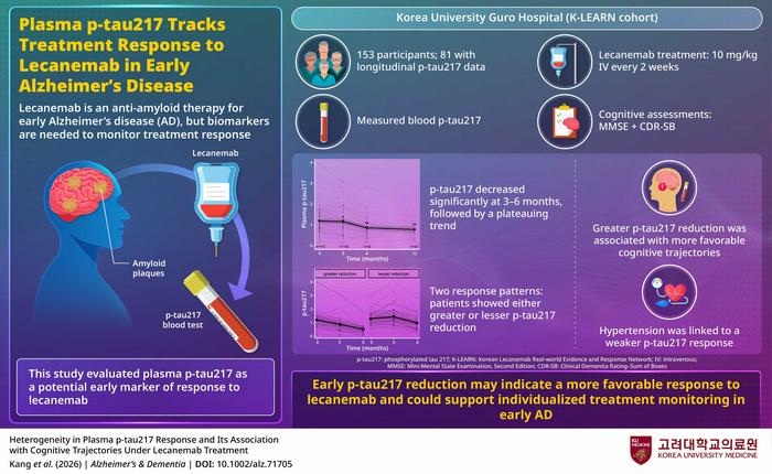
- Blood Biomarker May Help Monitor Early Response to Lecanemab in Alzheimer’s Disease
- Blood Antibody Linked to Lower Depression and PTSD Symptoms After Brain Injury
- Childhood Blood Biomarkers May Reveal Early Heart, Kidney, and Metabolic Risks
- Blood Biomarker Study Reveals Early Signals of Future Cognitive Difficulties
- Combined Blood and Genetic Tests Estimate Timing of Alzheimer’s Symptom Onset
- Blood Protein Analysis Helps Interpret Uncertain Rare Disease Variants
- 40-Gene Test Validated for High-Risk Squamous Cell Carcinoma
- Common Genetic Marker Predicts Faster Motor Decline in Parkinson’s Disease
- Blood Test Helps Clarify Prostate Cancer Risk After Elevated PSA
- Borderline Anemia May Carry Higher Risk Than Current Thresholds Suggest
- Biomarker-Guided Framework Aims to Guide Mantle Cell Lymphoma Therapy
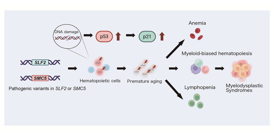
- New Genetic Findings Reveal Cause of Bone Marrow Failure Syndrome
- Ultra-Portable Device Enables Finger-Prick Blood Testing at Home
- Age-Specific CBC Reference Intervals Support Pediatric Diagnosis in Vietnam
- Temporal Immune Profiling Reveals How Sepsis States Change Over Time
- Study Identifies Immune Cells That Drive Harmful Autoantibody Responses in COVID-19
- Gut “Memory” May Explain Why IBD Flares Return
- AI-Based Antibody Profiling Predicts Strength of COVID-19 Vaccine Response
- Immune Biomarkers May Predict Recurrent Checkpoint Inhibitor Arthritis
- Infant Skin Microbiome Show Links to Later Eczema and Food Allergies
- Diagnostic Stewardship Guide Targets Testing Overuse and Hospital Cost Pressures
- Research Strengthens Bundibugyo Virus Outbreak Readiness with Faster Diagnostics
- Gut Microbiome Classifier Improves Colorectal Cancer Risk Stratification
- New Diagnostic Workflow Identifies Bloodstream Pathogens and Antibiotic Response in Hours
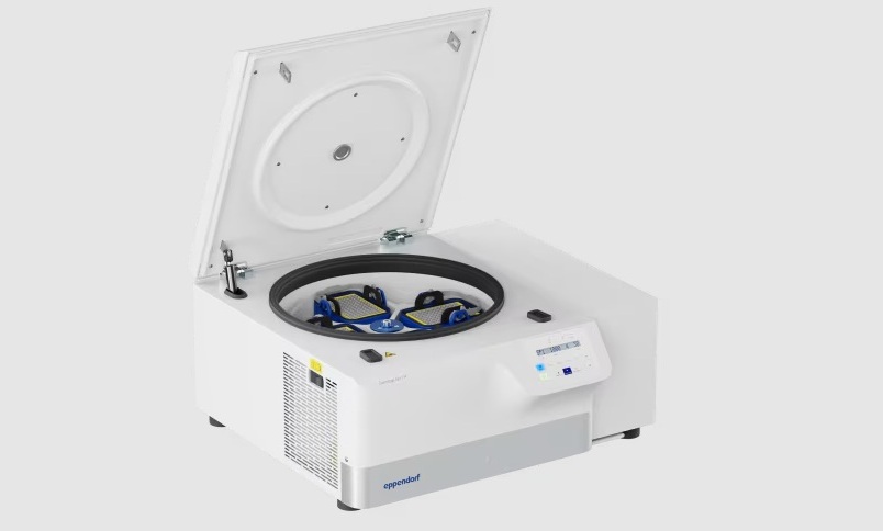
- New Multipurpose Centrifuge Combines High Capacity with Sustainable Cooling
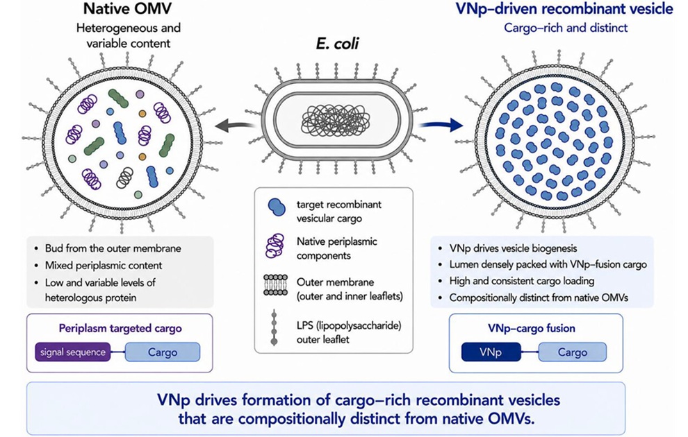
- Bacterial Vesicle Expression System Streamlines Production of Cancer Diagnostic Proteins
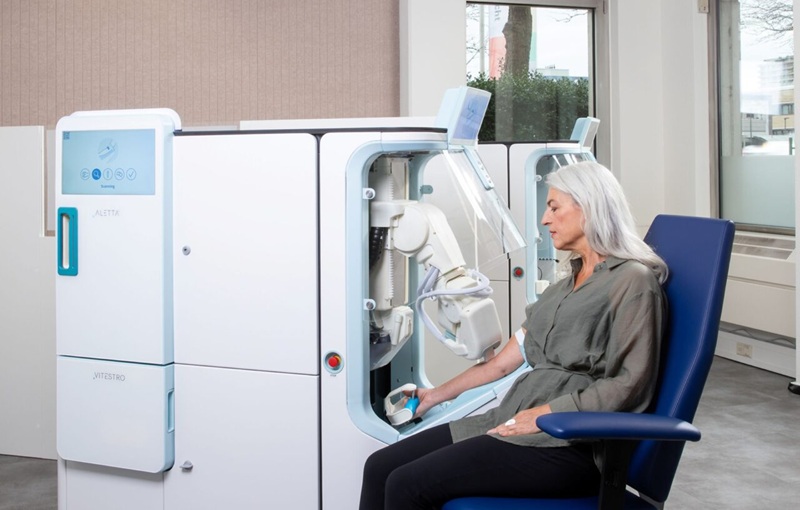
- Autonomous Robotic System Receives FDA Authorization for Blood Collection
- Interoperable Data Platform Standardizes Multi-Cancer Blood Test Results
- Training Device Improves Accuracy of Pooled Molecular Diagnostics
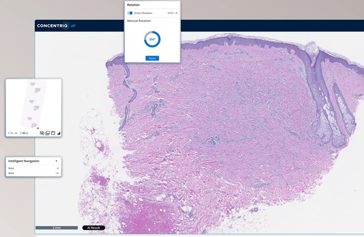
- Unilabs Expands Digital Pathology Across European Network with Proscia Platform
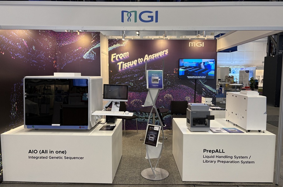
- MGI Launches Automated Multiplex Tissue Staining and Imaging System at ECP 2026
- Digital Pathology Partnership Integrates Breast Cancer Diagnostic and Prognostic Tools
- Blood-Based Neurology Biomarker Assays Authorized for Use in Brazil
- Sepsis Diagnostics Market Growth Fueled by Faster Pathogen Detection Needs
- New Gene-Disease Link May Help Diagnose Rare Neurodevelopmental Disorders
- Researchers Identify Shared Molecular Networks Behind Fatigue-Related Illnesses
- New Immune Target Could Support More Precise High Blood Pressure Treatment
- Whole-Blood Extracellular Vesicle Analysis Captures Molecular Signals Missed by Plasma Testing
- Blood Metabolite Signature Predicts ALS Progression and Points to Treatment Strategy
- Hybrid Computational Imaging Method Improves Digital Pathology Resolution
- FDA Clearance Expands Digital Pathology Options for Clinical Laboratories
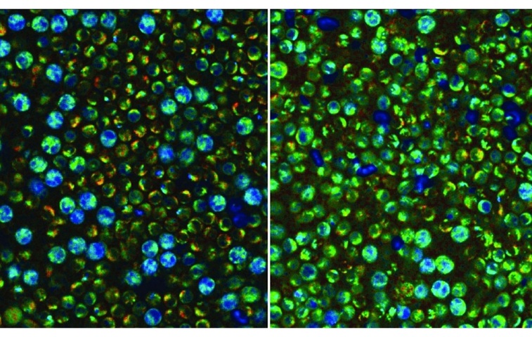
- Label-Free Imaging Reveals Hidden Immune Activity in Blood Samples
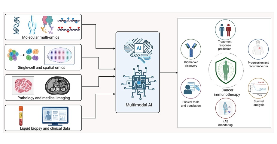
- AI Tool Improves Prediction of Immunotherapy Response in Lung Cancer
- New Journal Addresses Evolving Needs in Clinical and Translational Pathology
 Expo
Expo
- Machine Learning Supports Targeted Screening for Elevated Lipoprotein(a)
- Blood Biomarker May Help Monitor Early Response to Lecanemab in Alzheimer’s Disease
- Blood Antibody Linked to Lower Depression and PTSD Symptoms After Brain Injury
- Childhood Blood Biomarkers May Reveal Early Heart, Kidney, and Metabolic Risks
- Blood Biomarker Study Reveals Early Signals of Future Cognitive Difficulties
- Combined Blood and Genetic Tests Estimate Timing of Alzheimer’s Symptom Onset
- Blood Protein Analysis Helps Interpret Uncertain Rare Disease Variants
- 40-Gene Test Validated for High-Risk Squamous Cell Carcinoma
- Common Genetic Marker Predicts Faster Motor Decline in Parkinson’s Disease
- Blood Test Helps Clarify Prostate Cancer Risk After Elevated PSA
- Borderline Anemia May Carry Higher Risk Than Current Thresholds Suggest
- Biomarker-Guided Framework Aims to Guide Mantle Cell Lymphoma Therapy
- New Genetic Findings Reveal Cause of Bone Marrow Failure Syndrome
- Ultra-Portable Device Enables Finger-Prick Blood Testing at Home
- Age-Specific CBC Reference Intervals Support Pediatric Diagnosis in Vietnam
- Temporal Immune Profiling Reveals How Sepsis States Change Over Time
- Study Identifies Immune Cells That Drive Harmful Autoantibody Responses in COVID-19
- Gut “Memory” May Explain Why IBD Flares Return
- AI-Based Antibody Profiling Predicts Strength of COVID-19 Vaccine Response
- Immune Biomarkers May Predict Recurrent Checkpoint Inhibitor Arthritis
- Infant Skin Microbiome Show Links to Later Eczema and Food Allergies
- Diagnostic Stewardship Guide Targets Testing Overuse and Hospital Cost Pressures
- Research Strengthens Bundibugyo Virus Outbreak Readiness with Faster Diagnostics
- Gut Microbiome Classifier Improves Colorectal Cancer Risk Stratification
- New Diagnostic Workflow Identifies Bloodstream Pathogens and Antibiotic Response in Hours
- New Multipurpose Centrifuge Combines High Capacity with Sustainable Cooling
- Bacterial Vesicle Expression System Streamlines Production of Cancer Diagnostic Proteins
- Autonomous Robotic System Receives FDA Authorization for Blood Collection
- Interoperable Data Platform Standardizes Multi-Cancer Blood Test Results
- Training Device Improves Accuracy of Pooled Molecular Diagnostics
- Unilabs Expands Digital Pathology Across European Network with Proscia Platform
- MGI Launches Automated Multiplex Tissue Staining and Imaging System at ECP 2026
- Digital Pathology Partnership Integrates Breast Cancer Diagnostic and Prognostic Tools
- Blood-Based Neurology Biomarker Assays Authorized for Use in Brazil
- Sepsis Diagnostics Market Growth Fueled by Faster Pathogen Detection Needs
- New Gene-Disease Link May Help Diagnose Rare Neurodevelopmental Disorders
- Researchers Identify Shared Molecular Networks Behind Fatigue-Related Illnesses
- New Immune Target Could Support More Precise High Blood Pressure Treatment
- Whole-Blood Extracellular Vesicle Analysis Captures Molecular Signals Missed by Plasma Testing
- Blood Metabolite Signature Predicts ALS Progression and Points to Treatment Strategy
- Hybrid Computational Imaging Method Improves Digital Pathology Resolution
- FDA Clearance Expands Digital Pathology Options for Clinical Laboratories
- Label-Free Imaging Reveals Hidden Immune Activity in Blood Samples
- AI Tool Improves Prediction of Immunotherapy Response in Lung Cancer
- New Journal Addresses Evolving Needs in Clinical and Translational Pathology
































 Client Login