




Expo
view channel
view channel
view channel
view channel
view channel
view channel
view channel
view channel
view channel
Clinical Chem.Molecular DiagnosticsHematologyImmunologyMicrobiology
TechnologyIndustry
Events
Webinars

- Head-to-Head Study Compares Blood Tests for Early Alzheimer’s Disease Detection
- Mathematical Biomarkers Use Routine Blood Tests to Guide Adaptive Prostate Cancer Therapy
- Blood Metabolite Patterns May Enable Early Detection of Blood-Brain Barrier Injury
- Blood Biomarker Study Reveals Population-Specific Differences in Alzheimer’s Disease
- New Chairside Oral Swab Test Could Guide Biopsy Decisions in Oral Cancer
- Blood Test for Lung Cancer Screening Receives FDA Breakthrough Device Designation
- Sequencing-Based Newborn Screening Identifies Pediatric Cancer Risk at Birth
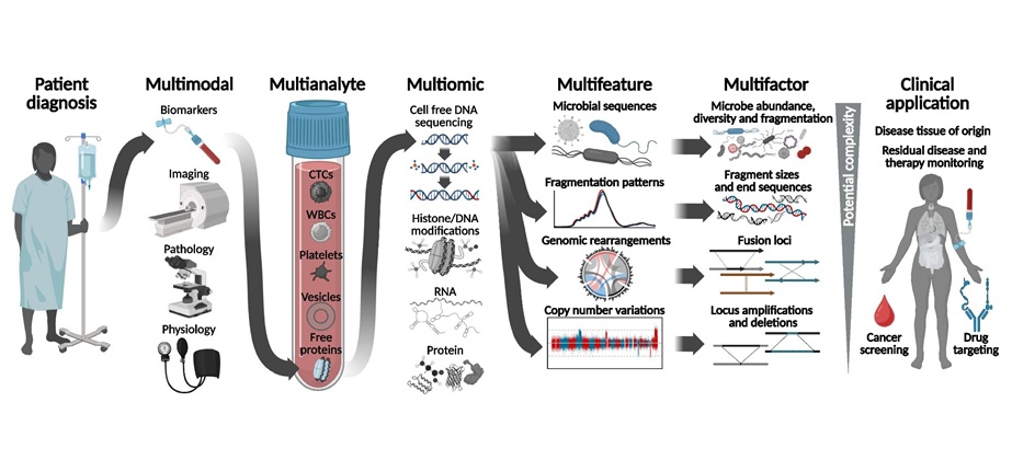
- Liquid Biopsy Combines Multiple Signals for Cancer Detection in a Single Analysis
- Newborn Screening Program Adds Spinal Muscular Atrophy Testing Across England
- Whole-Cell Genomic Analysis Expands the Potential of Liquid Biopsy
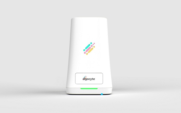
- Ultra-Portable Device Enables Finger-Prick Blood Testing at Home
- Age-Specific CBC Reference Intervals Support Pediatric Diagnosis in Vietnam
- Spectral Flow Cytometry Assay Enhances MRD Detection in Multiple Myeloma
- New Marker Helps Detect Aggressive Multiple Myeloma Earlier
- New Biomarkers Predict Resistance to Targeted Therapy in Rare Blood Cancer
- Immune Cell Blood Test May Predict Melanoma Immunotherapy Response
- Study Reveals Viral Protein Driving COVID-19-Related Vascular Injury
- Study Reveals Immune Mechanism Driving Severe COVID-19 Progression
- Antibody Profiling Identifies Preclinical Inflammatory Bowel Disease Years Before Diagnosis
- Ultrasensitive Blood Test Detects Sjögren’s Signature Years Before Diagnosis
- Plasma Cell-Free DNA Test Enables Earlier Diagnosis of Invasive Fungal Infections
- FDA Clears One-Hour Panel for Bloodstream Infection and AMR Detection
- Precision Screening Helps Close Hepatitis C Diagnosis and Treatment Gaps
- Proteomic Workflow Maps Microbial and Host Responses in Intestinal Inflammation
- Metagenomic Sequencing Enables Faster Diagnosis of Respiratory Infections in Cystic Fibrosis
- Training Device Improves Accuracy of Pooled Molecular Diagnostics
- New CE-Certified Software Advances Whole-Genome Cancer Testing
- National Rare Disease Registry Standardizes Genetic and Clinical Data for Coordinated Care
- AI Platform Links Biomarker Results to Cancer Clinical Trials and Guidelines
- Agentic AI Platform Supports Genomic Decision-Making in Oncology
- Collaboration Supports Global ADC Development with AI-Powered Tissue Analysis
- Standardized Staining Technology Advances Digital Pathology Workflows
- Global Testing Service Advances Leukemia MRD Monitoring
- Ultrasensitive MRD Assay Gains Medicare Coverage for Immunotherapy and Breast Cancer Monitoring
- Genprex and Roche Collaborate on AI-Driven NSCLC Biomarker Testing
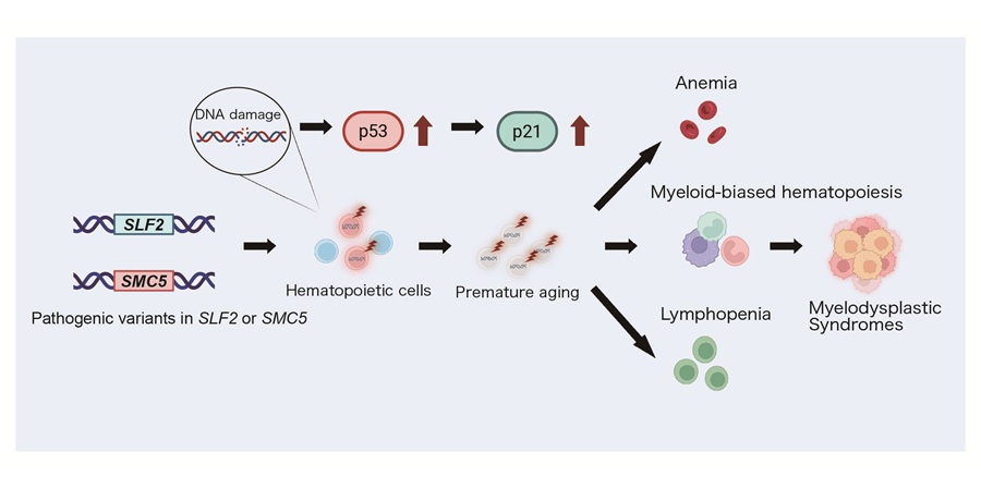
- New Genetic Cause Identified for Neurodevelopmental Disorder
- New Genetic Discovery Could Support Precision Diabetes Care
- Inherited Genetic Differences Help Explain Variable CAR T-Cell Therapy Outcomes
- AI-Powered Genome Mapping Reveals New Layer of Alzheimer’s Disease Biology
- Genetic Variations Reveal Mechanisms Behind Sudden Cardiac Death Risk
- AI Tool Identifies Slide Artifacts to Speed Digital Pathology Diagnosis
- FDA-Cleared Digital Pathology Platform Expands Interoperability for Primary Diagnosis
- AI Pathology Tool Predicts Relapse Risk in Stage II Colorectal Cancer
- AI Pathology Tool Stratifies Rectal Cancer to Guide Chemoradiotherapy
- PD-L1 Assay Guides Pembrolizumab Eligibility in Ovarian, Fallopian Tube, and Peritoneal Cancers
 Expo
Expo
- Head-to-Head Study Compares Blood Tests for Early Alzheimer’s Disease Detection
- Mathematical Biomarkers Use Routine Blood Tests to Guide Adaptive Prostate Cancer Therapy
- Blood Metabolite Patterns May Enable Early Detection of Blood-Brain Barrier Injury
- Blood Biomarker Study Reveals Population-Specific Differences in Alzheimer’s Disease
- New Chairside Oral Swab Test Could Guide Biopsy Decisions in Oral Cancer
- Blood Test for Lung Cancer Screening Receives FDA Breakthrough Device Designation
- Sequencing-Based Newborn Screening Identifies Pediatric Cancer Risk at Birth
- Liquid Biopsy Combines Multiple Signals for Cancer Detection in a Single Analysis
- Newborn Screening Program Adds Spinal Muscular Atrophy Testing Across England
- Whole-Cell Genomic Analysis Expands the Potential of Liquid Biopsy
- Ultra-Portable Device Enables Finger-Prick Blood Testing at Home
- Age-Specific CBC Reference Intervals Support Pediatric Diagnosis in Vietnam
- Spectral Flow Cytometry Assay Enhances MRD Detection in Multiple Myeloma
- New Marker Helps Detect Aggressive Multiple Myeloma Earlier
- New Biomarkers Predict Resistance to Targeted Therapy in Rare Blood Cancer
- Immune Cell Blood Test May Predict Melanoma Immunotherapy Response
- Study Reveals Viral Protein Driving COVID-19-Related Vascular Injury
- Study Reveals Immune Mechanism Driving Severe COVID-19 Progression
- Antibody Profiling Identifies Preclinical Inflammatory Bowel Disease Years Before Diagnosis
- Ultrasensitive Blood Test Detects Sjögren’s Signature Years Before Diagnosis
- Plasma Cell-Free DNA Test Enables Earlier Diagnosis of Invasive Fungal Infections
- FDA Clears One-Hour Panel for Bloodstream Infection and AMR Detection
- Precision Screening Helps Close Hepatitis C Diagnosis and Treatment Gaps
- Proteomic Workflow Maps Microbial and Host Responses in Intestinal Inflammation
- Metagenomic Sequencing Enables Faster Diagnosis of Respiratory Infections in Cystic Fibrosis
- Training Device Improves Accuracy of Pooled Molecular Diagnostics
- New CE-Certified Software Advances Whole-Genome Cancer Testing
- National Rare Disease Registry Standardizes Genetic and Clinical Data for Coordinated Care
- AI Platform Links Biomarker Results to Cancer Clinical Trials and Guidelines
- Agentic AI Platform Supports Genomic Decision-Making in Oncology
- Collaboration Supports Global ADC Development with AI-Powered Tissue Analysis
- Standardized Staining Technology Advances Digital Pathology Workflows
- Global Testing Service Advances Leukemia MRD Monitoring
- Ultrasensitive MRD Assay Gains Medicare Coverage for Immunotherapy and Breast Cancer Monitoring
- Genprex and Roche Collaborate on AI-Driven NSCLC Biomarker Testing
- New Genetic Cause Identified for Neurodevelopmental Disorder
- New Genetic Discovery Could Support Precision Diabetes Care
- Inherited Genetic Differences Help Explain Variable CAR T-Cell Therapy Outcomes
- AI-Powered Genome Mapping Reveals New Layer of Alzheimer’s Disease Biology
- Genetic Variations Reveal Mechanisms Behind Sudden Cardiac Death Risk
- AI Tool Identifies Slide Artifacts to Speed Digital Pathology Diagnosis
- FDA-Cleared Digital Pathology Platform Expands Interoperability for Primary Diagnosis
- AI Pathology Tool Predicts Relapse Risk in Stage II Colorectal Cancer
- AI Pathology Tool Stratifies Rectal Cancer to Guide Chemoradiotherapy
- PD-L1 Assay Guides Pembrolizumab Eligibility in Ovarian, Fallopian Tube, and Peritoneal Cancers


































 Client Login