




Expo
view channel
view channel
view channel
view channel
view channel
view channel
view channel
view channel
view channel
Clinical Chem.Molecular DiagnosticsHematologyImmunologyMicrobiology
TechnologyIndustry
Events
Webinars

- Blood-Based Score May Support Targeted Interventions for Healthier Aging
- Noninvasive Tests Offer Alternatives to Biopsy for Assessing Advanced Liver Scarring
- Blood Proteins Linked to Mortality Risk and Biological Aging Across Organ Systems
- Two-Step Blood Test Improves Specificity for Alzheimer’s Diagnosis
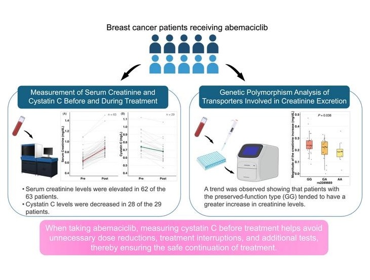
- Common Kidney Function Tests May Miss CKD in Cancer Patients
- Blood Test Identifies Early Breast and Prostate Cancer Signals Years Before Diagnosis
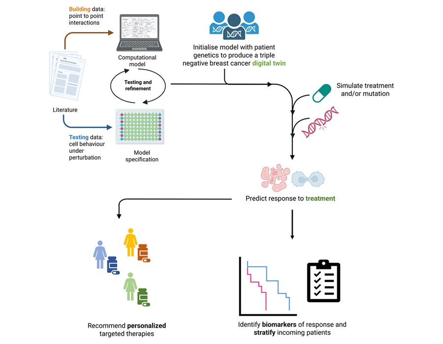
- Digital Twins Predict Treatment Response in Triple-Negative Breast Cancer
- Tumor and Immune Biomarkers Linked to Treatment Response in T-Cell Lymphoma
- Blood MRD Testing Aids Risk Analysis in Bladder and Other Cancers
- Liquid Biopsy Measures Tumor Burden and Identifies Small Cell Lung Cancer Subtypes
- New Risk-Stratification Tool Predicts Relapse After Blinatumomab in Childhood ALL
- New Myelofibrosis Guidelines Recommend Expanded Genetic Testing at Diagnosis
- Handheld Point-of-Care Test Aims to Enable Rapid Coagulation Assessment in Trauma
- New Donor Genetic Marker May Help Predict Stem Cell Transplant Success
- Updated Ferritin Thresholds Improve Detection of Iron Deficiency
- Prototype Point-of-Care Lateral Flow Test Aims to Improve Neonatal Sepsis Triage
- Immune and Inflammatory Markers May Help Assess CAR T-Cell Complication Risk
- COVID-19 and Flu Combo Test Launches With FDA Clearance
- Compact Immunoanalyzer Gains CE Mark for Testing Across Local Care Settings
- Antibody Profiling Predicts Inflammatory Bowel Disease Years Before Diagnosis
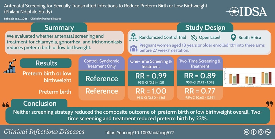
- Repeat Prenatal STI Screening and Treatment May Reduce Preterm Births
- Decentralized Laboratory Network Improves Detection of Viral Hemorrhagic Fevers in Guinea
- Molecular Panel Gains WHO Emergency Use Listing to Support Ebola Outbreak Response
- Rapid Host-Response Test Aims to Predict Corticosteroid Benefit in Sepsis
- Oral and Lung Microbiome Changes Linked to Severe Long Covid Symptoms
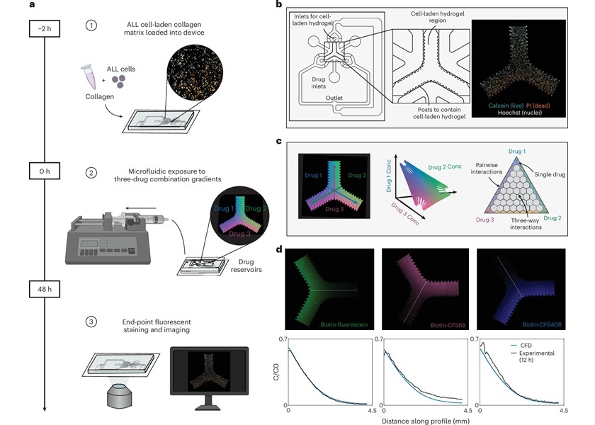
- Microfluidic Assay Predicts Treatment Response in Pediatric Leukemia
- AI Models Use Routine Blood Tests and Health Records to Predict Cancer Risk
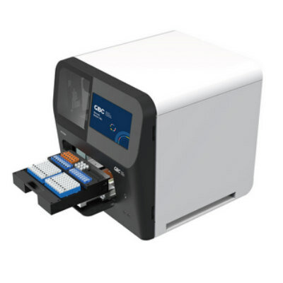
- Compact PCR Thermal Cycler Launches for Routine Molecular Laboratory Workflows
- New AI-Powered System Aims to Streamline Laboratory and Manufacturing Workflows
- Laser-Based Swab Analysis Shows Promise for Detecting Disease-Linked Odor Patterns
- CE-Marked IVD Sequencing Platform Launches Across Ten EU Countries
- Collaboration Brings Tumor-Informed MRD Monitoring to Breast Cancer Care in Abu Dhabi
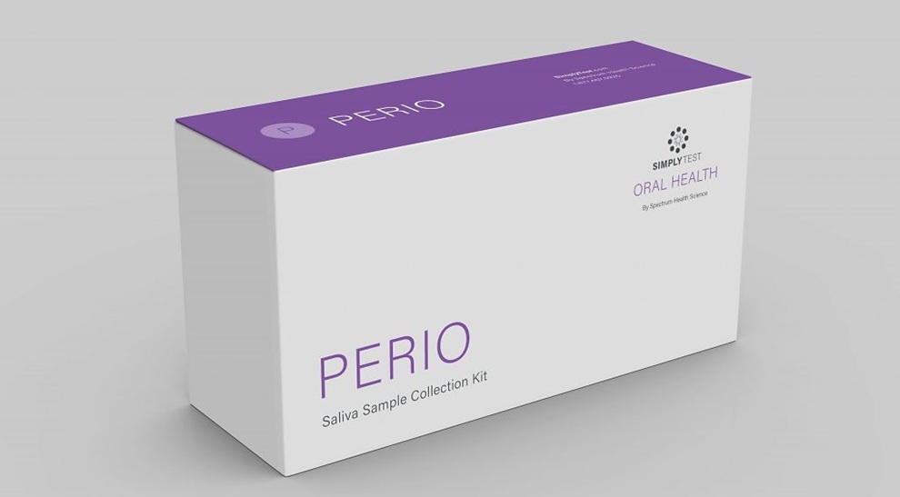
- Real Diagnostics Adds Saliva-Based Periodontal Testing With SimplyTest Deal
- Qiagen Agrees to Go Private Through EQT-Led Acquisition
- Hologic and Inpeco Partner to Advance Molecular Laboratory Automation in Europe
- Lung Tumor RNA Variants Offer Clues to Immunotherapy Benefit
- Population-Scale Genome Survey Supports Interpretation of Genetic Testing
- Single-Cell Profiling Reveals Chemotherapy Resistance Signals in Ovarian Cancer
- Genomic Tool Supports Analysis of Repeat Expansions Linked to Inherited Disorders
- CSF Biomarker Meprin-Beta Increases in Alzheimer’s Disease
- Breakthrough-Designated Cytology Test Supports Oral Cancer Risk Assessment
- AI Pathology Approach Closely Aligns With Breast Cancer Genomic Risk Classification
- Roche Launches Automated Test to Support Mesothelioma Differential Diagnosis
- Digital Pathology Test Stratifies Recurrence Risk in Early Breast Cancer
- AI Pathology Tool Predicts DNA Repair Deficiency in High-Grade Serous Ovarian Cancer
 Expo
- Blood-Based Score May Support Targeted Interventions for Healthier Aging
- Noninvasive Tests Offer Alternatives to Biopsy for Assessing Advanced Liver Scarring
- Blood Proteins Linked to Mortality Risk and Biological Aging Across Organ Systems
- Two-Step Blood Test Improves Specificity for Alzheimer’s Diagnosis
- Common Kidney Function Tests May Miss CKD in Cancer Patients
- Blood Test Identifies Early Breast and Prostate Cancer Signals Years Before Diagnosis
- Digital Twins Predict Treatment Response in Triple-Negative Breast Cancer
- Tumor and Immune Biomarkers Linked to Treatment Response in T-Cell Lymphoma
- Blood MRD Testing Aids Risk Analysis in Bladder and Other Cancers
- Liquid Biopsy Measures Tumor Burden and Identifies Small Cell Lung Cancer Subtypes
- New Risk-Stratification Tool Predicts Relapse After Blinatumomab in Childhood ALL
- New Myelofibrosis Guidelines Recommend Expanded Genetic Testing at Diagnosis
- Handheld Point-of-Care Test Aims to Enable Rapid Coagulation Assessment in Trauma
- New Donor Genetic Marker May Help Predict Stem Cell Transplant Success
- Updated Ferritin Thresholds Improve Detection of Iron Deficiency
- Prototype Point-of-Care Lateral Flow Test Aims to Improve Neonatal Sepsis Triage
- Immune and Inflammatory Markers May Help Assess CAR T-Cell Complication Risk
- COVID-19 and Flu Combo Test Launches With FDA Clearance
- Compact Immunoanalyzer Gains CE Mark for Testing Across Local Care Settings
- Antibody Profiling Predicts Inflammatory Bowel Disease Years Before Diagnosis
- Repeat Prenatal STI Screening and Treatment May Reduce Preterm Births
- Decentralized Laboratory Network Improves Detection of Viral Hemorrhagic Fevers in Guinea
- Molecular Panel Gains WHO Emergency Use Listing to Support Ebola Outbreak Response
- Rapid Host-Response Test Aims to Predict Corticosteroid Benefit in Sepsis
- Oral and Lung Microbiome Changes Linked to Severe Long Covid Symptoms
- Microfluidic Assay Predicts Treatment Response in Pediatric Leukemia
- AI Models Use Routine Blood Tests and Health Records to Predict Cancer Risk
- Compact PCR Thermal Cycler Launches for Routine Molecular Laboratory Workflows
- New AI-Powered System Aims to Streamline Laboratory and Manufacturing Workflows
- Laser-Based Swab Analysis Shows Promise for Detecting Disease-Linked Odor Patterns
- CE-Marked IVD Sequencing Platform Launches Across Ten EU Countries
- Collaboration Brings Tumor-Informed MRD Monitoring to Breast Cancer Care in Abu Dhabi
- Real Diagnostics Adds Saliva-Based Periodontal Testing With SimplyTest Deal
- Qiagen Agrees to Go Private Through EQT-Led Acquisition
- Hologic and Inpeco Partner to Advance Molecular Laboratory Automation in Europe
- Lung Tumor RNA Variants Offer Clues to Immunotherapy Benefit
- Population-Scale Genome Survey Supports Interpretation of Genetic Testing
- Single-Cell Profiling Reveals Chemotherapy Resistance Signals in Ovarian Cancer
- Genomic Tool Supports Analysis of Repeat Expansions Linked to Inherited Disorders
- CSF Biomarker Meprin-Beta Increases in Alzheimer’s Disease
- Breakthrough-Designated Cytology Test Supports Oral Cancer Risk Assessment
- AI Pathology Approach Closely Aligns With Breast Cancer Genomic Risk Classification
- Roche Launches Automated Test to Support Mesothelioma Differential Diagnosis
- Digital Pathology Test Stratifies Recurrence Risk in Early Breast Cancer
- AI Pathology Tool Predicts DNA Repair Deficiency in High-Grade Serous Ovarian Cancer






































 Client Login