





Expo
view channel
view channel
view channel
view channel
view channel
view channel
view channel
view channel
view channel
Clinical Chem.Molecular DiagnosticsHematologyImmunologyMicrobiology
TechnologyIndustry
Events
Webinars

- Multi-Biomarker Blood Test Shows Promise for Early Pancreatic Cancer Detection
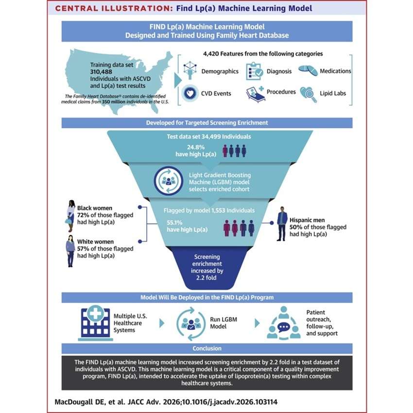
- Machine Learning Supports Targeted Screening for Elevated Lipoprotein(a)
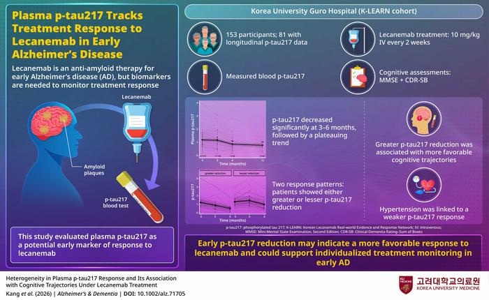
- Blood Biomarker May Help Monitor Early Response to Lecanemab in Alzheimer’s Disease
- Blood Antibody Linked to Lower Depression and PTSD Symptoms After Brain Injury
- Childhood Blood Biomarkers May Reveal Early Heart, Kidney, and Metabolic Risks
- FDA Clears Compact Molecular COVID Test for Decentralized Care Settings
- Blood-Based DNA Analysis Method Detects Early-Stage Solid Cancers
- Combined Blood and Genetic Tests Estimate Timing of Alzheimer’s Symptom Onset
- Blood Protein Analysis Helps Interpret Uncertain Rare Disease Variants
- 40-Gene Test Validated for High-Risk Squamous Cell Carcinoma
- New Automated Hemostasis Systems Become Available Across US Laboratories
- Variation in AML Residual Disease Testing Prompts Push for Standardization
- Hidden Molecular Mechanism Behind HIT Blood-Clotting Disorder Revealed
- Borderline Anemia May Carry Higher Risk Than Current Thresholds Suggest
- Biomarker-Guided Framework Aims to Guide Mantle Cell Lymphoma Therapy
- New Cellular Map May Help Predict Crohn’s Disease Course in Children
- Temporal Immune Profiling Reveals How Sepsis States Change Over Time
- Study Identifies Immune Cells That Drive Harmful Autoantibody Responses in COVID-19
- Gut “Memory” May Explain Why IBD Flares Return
- AI-Based Antibody Profiling Predicts Strength of COVID-19 Vaccine Response
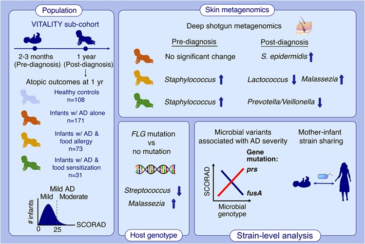
- Infant Skin Microbiome Show Links to Later Eczema and Food Allergies
- Diagnostic Stewardship Guide Targets Testing Overuse and Hospital Cost Pressures
- Research Strengthens Bundibugyo Virus Outbreak Readiness with Faster Diagnostics
- Gut Microbiome Classifier Improves Colorectal Cancer Risk Stratification
- New Diagnostic Workflow Identifies Bloodstream Pathogens and Antibiotic Response in Hours
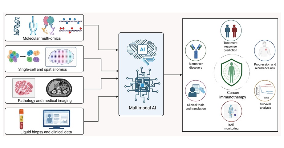
- Multimodal AI Framework Aims to Guide Cancer Immunotherapy Decisions
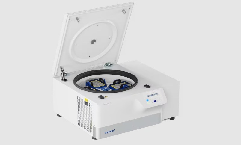
- New Multipurpose Centrifuge Combines High Capacity with Sustainable Cooling
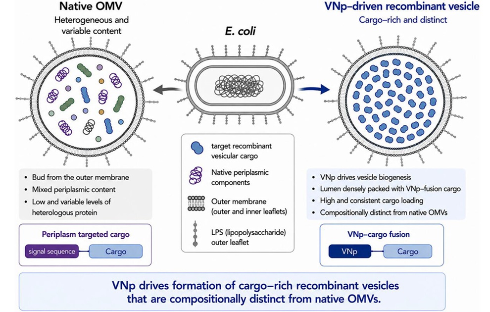
- Bacterial Vesicle Expression System Streamlines Production of Cancer Diagnostic Proteins
- Autonomous Robotic System Receives FDA Authorization for Blood Collection
- Interoperable Data Platform Standardizes Multi-Cancer Blood Test Results
- Unilabs Expands Digital Pathology Across European Network with Proscia Platform
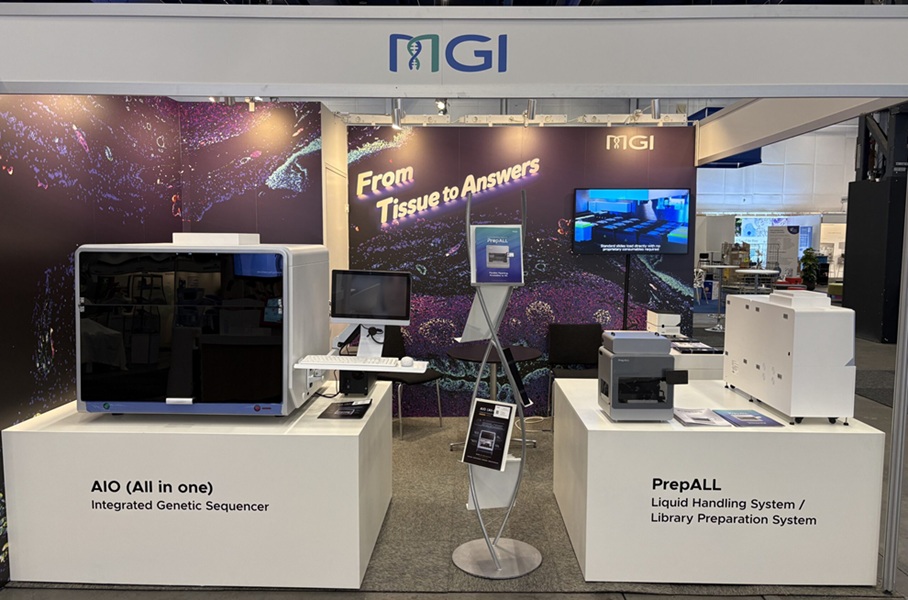
- MGI Launches Automated Multiplex Tissue Staining and Imaging System at ECP 2026
- Digital Pathology Partnership Integrates Breast Cancer Diagnostic and Prognostic Tools
- Blood-Based Neurology Biomarker Assays Authorized for Use in Brazil
- Sepsis Diagnostics Market Growth Fueled by Faster Pathogen Detection Needs
- New Autoantibody Target Identified in Neuromyelitis Optica Spectrum Disorder
- New Gene-Disease Link May Help Diagnose Rare Neurodevelopmental Disorders
- Researchers Identify Shared Molecular Networks Behind Fatigue-Related Illnesses
- New Immune Target Could Support More Precise High Blood Pressure Treatment
- Whole-Blood Extracellular Vesicle Analysis Captures Molecular Signals Missed by Plasma Testing
- Tumor Budding Grading May Predict Chemotherapy Benefit in Resected Lung Squamous Cancer
- CAP Updates Cancer Protocols to Refine Breast Cancer Biomarker Reporting and GIST Molecular Testing
- AI Spatial Analysis of Routine Slides Helps Predict Pancreatic Cancer Recurrence
- Hybrid Computational Imaging Method Improves Digital Pathology Resolution
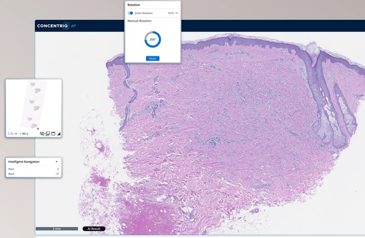
- FDA Clearance Expands Digital Pathology Options for Clinical Laboratories
 Expo
Expo
- Multi-Biomarker Blood Test Shows Promise for Early Pancreatic Cancer Detection
- Machine Learning Supports Targeted Screening for Elevated Lipoprotein(a)
- Blood Biomarker May Help Monitor Early Response to Lecanemab in Alzheimer’s Disease
- Blood Antibody Linked to Lower Depression and PTSD Symptoms After Brain Injury
- Childhood Blood Biomarkers May Reveal Early Heart, Kidney, and Metabolic Risks
- FDA Clears Compact Molecular COVID Test for Decentralized Care Settings
- Blood-Based DNA Analysis Method Detects Early-Stage Solid Cancers
- Combined Blood and Genetic Tests Estimate Timing of Alzheimer’s Symptom Onset
- Blood Protein Analysis Helps Interpret Uncertain Rare Disease Variants
- 40-Gene Test Validated for High-Risk Squamous Cell Carcinoma
- New Automated Hemostasis Systems Become Available Across US Laboratories
- Variation in AML Residual Disease Testing Prompts Push for Standardization
- Hidden Molecular Mechanism Behind HIT Blood-Clotting Disorder Revealed
- Borderline Anemia May Carry Higher Risk Than Current Thresholds Suggest
- Biomarker-Guided Framework Aims to Guide Mantle Cell Lymphoma Therapy
- New Cellular Map May Help Predict Crohn’s Disease Course in Children
- Temporal Immune Profiling Reveals How Sepsis States Change Over Time
- Study Identifies Immune Cells That Drive Harmful Autoantibody Responses in COVID-19
- Gut “Memory” May Explain Why IBD Flares Return
- AI-Based Antibody Profiling Predicts Strength of COVID-19 Vaccine Response
- Infant Skin Microbiome Show Links to Later Eczema and Food Allergies
- Diagnostic Stewardship Guide Targets Testing Overuse and Hospital Cost Pressures
- Research Strengthens Bundibugyo Virus Outbreak Readiness with Faster Diagnostics
- Gut Microbiome Classifier Improves Colorectal Cancer Risk Stratification
- New Diagnostic Workflow Identifies Bloodstream Pathogens and Antibiotic Response in Hours
- Multimodal AI Framework Aims to Guide Cancer Immunotherapy Decisions
- New Multipurpose Centrifuge Combines High Capacity with Sustainable Cooling
- Bacterial Vesicle Expression System Streamlines Production of Cancer Diagnostic Proteins
- Autonomous Robotic System Receives FDA Authorization for Blood Collection
- Interoperable Data Platform Standardizes Multi-Cancer Blood Test Results
- Unilabs Expands Digital Pathology Across European Network with Proscia Platform
- MGI Launches Automated Multiplex Tissue Staining and Imaging System at ECP 2026
- Digital Pathology Partnership Integrates Breast Cancer Diagnostic and Prognostic Tools
- Blood-Based Neurology Biomarker Assays Authorized for Use in Brazil
- Sepsis Diagnostics Market Growth Fueled by Faster Pathogen Detection Needs
- New Autoantibody Target Identified in Neuromyelitis Optica Spectrum Disorder
- New Gene-Disease Link May Help Diagnose Rare Neurodevelopmental Disorders
- Researchers Identify Shared Molecular Networks Behind Fatigue-Related Illnesses
- New Immune Target Could Support More Precise High Blood Pressure Treatment
- Whole-Blood Extracellular Vesicle Analysis Captures Molecular Signals Missed by Plasma Testing
- Tumor Budding Grading May Predict Chemotherapy Benefit in Resected Lung Squamous Cancer
- CAP Updates Cancer Protocols to Refine Breast Cancer Biomarker Reporting and GIST Molecular Testing
- AI Spatial Analysis of Routine Slides Helps Predict Pancreatic Cancer Recurrence
- Hybrid Computational Imaging Method Improves Digital Pathology Resolution
- FDA Clearance Expands Digital Pathology Options for Clinical Laboratories


































 Client Login